用于原位肝脏模型的核磁共振成像共聚焦啮齿动物组织切片阵列

IF 3 2区 工程技术 Q1 ACOUSTICS
Ryan Hubbard, David Choi, Tejaswi Worlikar, Ulrich Scheven, Hanna Kim, Jonathan Sukovich, Timothy L Hall, Zhen Xu
{"title":"用于原位肝脏模型的核磁共振成像共聚焦啮齿动物组织切片阵列","authors":"Ryan Hubbard, David Choi, Tejaswi Worlikar, Ulrich Scheven, Hanna Kim, Jonathan Sukovich, Timothy L Hall, Zhen Xu","doi":"10.1109/TUFFC.2025.3553083","DOIUrl":null,"url":null,"abstract":"<p><p>Histotripsy has emerged as a promising therapeutic option for liver tumors, recently gaining FDA approval for clinical use in October 2023. Preclinical in-vivo histotripsy experiments primarily utilize subcutaneous ectopic murine tumor models, which fail to accurately replicate the complex immunosuppressive tumor microenvironment (TME) of liver tumors. In order to address this gap, we present the design, development, and in-vivo demonstration of a miniature, electronically steerable MRI-guided histotripsy array tailored for orthotopic murine liver tumor models. This novel system integrates an 89-element phased array within a 7.0T small animal MRI scanner, enabling precise targeting through enhanced soft tissue contrast and three-dimensional visualization. The targeting accuracy of the array was validated in tissue-mimicking red blood cell phantoms, exhibiting targeting precision of 0.24 mm ± 0.1 mm. Subsequent in-vivo experiments in naïve mice demonstrated successful liver ablations, confirmed by gross morphology and histological analysis. However, the presence of grating lobes led to undesired collateral damage, highlighted by lung hemorrhages, necessitating future adjustments in the array's design. This study illustrates the foundational steps necessary for translating histotripsy experiments from subcutaneous to orthotopic models.</p>","PeriodicalId":13322,"journal":{"name":"IEEE transactions on ultrasonics, ferroelectrics, and frequency control","volume":"PP ","pages":""},"PeriodicalIF":3.0000,"publicationDate":"2025-03-20","publicationTypes":"Journal Article","fieldsOfStudy":null,"isOpenAccess":false,"openAccessPdf":"","citationCount":"0","resultStr":"{\"title\":\"MRI Co-registered Rodent Histotripsy Array for Orthotopic Liver Models.\",\"authors\":\"Ryan Hubbard, David Choi, Tejaswi Worlikar, Ulrich Scheven, Hanna Kim, Jonathan Sukovich, Timothy L Hall, Zhen Xu\",\"doi\":\"10.1109/TUFFC.2025.3553083\",\"DOIUrl\":null,\"url\":null,\"abstract\":\"<p><p>Histotripsy has emerged as a promising therapeutic option for liver tumors, recently gaining FDA approval for clinical use in October 2023. Preclinical in-vivo histotripsy experiments primarily utilize subcutaneous ectopic murine tumor models, which fail to accurately replicate the complex immunosuppressive tumor microenvironment (TME) of liver tumors. In order to address this gap, we present the design, development, and in-vivo demonstration of a miniature, electronically steerable MRI-guided histotripsy array tailored for orthotopic murine liver tumor models. This novel system integrates an 89-element phased array within a 7.0T small animal MRI scanner, enabling precise targeting through enhanced soft tissue contrast and three-dimensional visualization. The targeting accuracy of the array was validated in tissue-mimicking red blood cell phantoms, exhibiting targeting precision of 0.24 mm ± 0.1 mm. Subsequent in-vivo experiments in naïve mice demonstrated successful liver ablations, confirmed by gross morphology and histological analysis. However, the presence of grating lobes led to undesired collateral damage, highlighted by lung hemorrhages, necessitating future adjustments in the array's design. This study illustrates the foundational steps necessary for translating histotripsy experiments from subcutaneous to orthotopic models.</p>\",\"PeriodicalId\":13322,\"journal\":{\"name\":\"IEEE transactions on ultrasonics, ferroelectrics, and frequency control\",\"volume\":\"PP \",\"pages\":\"\"},\"PeriodicalIF\":3.0000,\"publicationDate\":\"2025-03-20\",\"publicationTypes\":\"Journal Article\",\"fieldsOfStudy\":null,\"isOpenAccess\":false,\"openAccessPdf\":\"\",\"citationCount\":\"0\",\"resultStr\":null,\"platform\":\"Semanticscholar\",\"paperid\":null,\"PeriodicalName\":\"IEEE transactions on ultrasonics, ferroelectrics, and frequency control\",\"FirstCategoryId\":\"5\",\"ListUrlMain\":\"https://doi.org/10.1109/TUFFC.2025.3553083\",\"RegionNum\":2,\"RegionCategory\":\"工程技术\",\"ArticlePicture\":[],\"TitleCN\":null,\"AbstractTextCN\":null,\"PMCID\":null,\"EPubDate\":\"\",\"PubModel\":\"\",\"JCR\":\"Q1\",\"JCRName\":\"ACOUSTICS\",\"Score\":null,\"Total\":0}","platform":"Semanticscholar","paperid":null,"PeriodicalName":"IEEE transactions on ultrasonics, ferroelectrics, and frequency control","FirstCategoryId":"5","ListUrlMain":"https://doi.org/10.1109/TUFFC.2025.3553083","RegionNum":2,"RegionCategory":"工程技术","ArticlePicture":[],"TitleCN":null,"AbstractTextCN":null,"PMCID":null,"EPubDate":"","PubModel":"","JCR":"Q1","JCRName":"ACOUSTICS","Score":null,"Total":0}
引用次数: 0

摘要

本文章由计算机程序翻译,如有差异,请以英文原文为准。
MRI Co-registered Rodent Histotripsy Array for Orthotopic Liver Models.

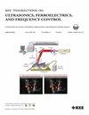
Histotripsy has emerged as a promising therapeutic option for liver tumors, recently gaining FDA approval for clinical use in October 2023. Preclinical in-vivo histotripsy experiments primarily utilize subcutaneous ectopic murine tumor models, which fail to accurately replicate the complex immunosuppressive tumor microenvironment (TME) of liver tumors. In order to address this gap, we present the design, development, and in-vivo demonstration of a miniature, electronically steerable MRI-guided histotripsy array tailored for orthotopic murine liver tumor models. This novel system integrates an 89-element phased array within a 7.0T small animal MRI scanner, enabling precise targeting through enhanced soft tissue contrast and three-dimensional visualization. The targeting accuracy of the array was validated in tissue-mimicking red blood cell phantoms, exhibiting targeting precision of 0.24 mm ± 0.1 mm. Subsequent in-vivo experiments in naïve mice demonstrated successful liver ablations, confirmed by gross morphology and histological analysis. However, the presence of grating lobes led to undesired collateral damage, highlighted by lung hemorrhages, necessitating future adjustments in the array's design. This study illustrates the foundational steps necessary for translating histotripsy experiments from subcutaneous to orthotopic models.

求助全文
通过发布文献求助,成功后即可免费获取论文全文。 去求助
来源期刊
CiteScore
7.70
自引率
16.70%
发文量
583
审稿时长
4.5 months
期刊介绍: IEEE Transactions on Ultrasonics, Ferroelectrics and Frequency Control includes the theory, technology, materials, and applications relating to: (1) the generation, transmission, and detection of ultrasonic waves and related phenomena; (2) medical ultrasound, including hyperthermia, bioeffects, tissue characterization and imaging; (3) ferroelectric, piezoelectric, and piezomagnetic materials, including crystals, polycrystalline solids, films, polymers, and composites; (4) frequency control, timing and time distribution, including crystal oscillators and other means of classical frequency control, and atomic, molecular and laser frequency control standards. Areas of interest range from fundamental studies to the design and/or applications of devices and systems.
×
引用
GB/T 7714-2015
复制
MLA
复制
APA
复制
导出至
BibTeX EndNote RefMan NoteFirst NoteExpress
×
提示
您的信息不完整,为了账户安全,请先补充。
现在去补充
×
提示
您因"违规操作"
具体请查看互助需知
我知道了
×
提示
确定
请完成安全验证×
copy
已复制链接
快去分享给好友吧!
我知道了
右上角分享
点击右上角分享
0
联系我们:info@booksci.cn Book学术提供免费学术资源搜索服务,方便国内外学者检索中英文文献。致力于提供最便捷和优质的服务体验。 Copyright © 2023 布克学术 All rights reserved.
京ICP备2023020795号-1
ghs 京公网安备 11010802042870号
Book学术文献互助
Book学术文献互助群
群 号:481959085
Book学术官方微信