Giovanni Marco Saladino, Dilyana B. Mangarova, Kerem Nernekli, Jie Wang, Giacomo Annio, Zahra Shokri Varniab, Zubeda Khatoon, Goreti Ribeiro Morais, Yifeng Shi, Edwin Chang, Laura J. Pisani, Grigory Tikhomirov, Robert A. Falconer and Heike E. Daldrup-Link
{"title":"利用磁共振成像和活体显微技术追踪胶质母细胞瘤中治疗性纳米颗粒积聚的多模态成像方法","authors":"Giovanni Marco Saladino, Dilyana B. Mangarova, Kerem Nernekli, Jie Wang, Giacomo Annio, Zahra Shokri Varniab, Zubeda Khatoon, Goreti Ribeiro Morais, Yifeng Shi, Edwin Chang, Laura J. Pisani, Grigory Tikhomirov, Robert A. Falconer and Heike E. Daldrup-Link","doi":"10.1039/D5NR00447K","DOIUrl":null,"url":null,"abstract":"<p >Theranostic nanoparticles (NPs) have been designed for simultaneous therapeutic and diagnostic purposes, thereby enabling personalized cancer therapy and <em>in vivo</em> drug tracking. However, studies thus far have focused on imaging NP tumor accumulation at the macroscopic level and correlating results with <em>ex vivo</em> histology. Limited evidence exists on whether <em>in vivo</em> NP tumor contrast enhancement on magnetic resonance imaging (MRI) correlates with <em>in vivo</em> NP tumor accumulation at the microscopic level. To address this gap, the purpose of our study was to correlate quantitative MRI estimates of NP accumulation with <em>in vivo</em> NP signal quantification as measured through two-photon intravital microscopy (IVM) in an orthotopic murine glioblastoma multiforme model (GBM). To enable multimodal imaging, we designed dual-mode NPs, composed of a carbohydrate-coated magnetic core (Ferumoxytol) as an MRI contrast agent, and a conjugated fluorophore (FITC) for IVM detection. We administered these NPs with or without a conjugated vascular disrupting agent (VDA) to assess its effect on NP delivery to GBM. We correlated <em>in vivo</em> MRI contrast enhancement in tumors, quantified as <em>T</em><small><sub>2</sub></small> relaxation time, with IVM fluorescence spatial decay rate. Results demonstrated a significantly lower tumor <em>T</em><small><sub>2</sub></small> relaxation time and spatial decay rate in tumors targeted with VDA-conjugated NPs compared to unconjugated NPs. <em>Postmortem</em> histological analyses validated the <em>in vivo</em> observations. The presented multimodal imaging approach enabled a quantitative correlation between MRI contrast enhancement at the macroscopic level and NP accumulation in the tumor microenvironment. These studies lay the groundwork for the precise evaluation of the tumor targeting of theranostic NPs.</p>","PeriodicalId":92,"journal":{"name":"Nanoscale","volume":" 16","pages":" 9986-9995"},"PeriodicalIF":5.1000,"publicationDate":"2025-03-19","publicationTypes":"Journal Article","fieldsOfStudy":null,"isOpenAccess":false,"openAccessPdf":"https://pubs.rsc.org/en/content/articlepdf/2025/nr/d5nr00447k?page=search","citationCount":"0","resultStr":"{\"title\":\"Multimodal imaging approach to track theranostic nanoparticle accumulation in glioblastoma with magnetic resonance imaging and intravital microscopy†\",\"authors\":\"Giovanni Marco Saladino, Dilyana B. Mangarova, Kerem Nernekli, Jie Wang, Giacomo Annio, Zahra Shokri Varniab, Zubeda Khatoon, Goreti Ribeiro Morais, Yifeng Shi, Edwin Chang, Laura J. Pisani, Grigory Tikhomirov, Robert A. Falconer and Heike E. Daldrup-Link\",\"doi\":\"10.1039/D5NR00447K\",\"DOIUrl\":null,\"url\":null,\"abstract\":\"<p >Theranostic nanoparticles (NPs) have been designed for simultaneous therapeutic and diagnostic purposes, thereby enabling personalized cancer therapy and <em>in vivo</em> drug tracking. However, studies thus far have focused on imaging NP tumor accumulation at the macroscopic level and correlating results with <em>ex vivo</em> histology. Limited evidence exists on whether <em>in vivo</em> NP tumor contrast enhancement on magnetic resonance imaging (MRI) correlates with <em>in vivo</em> NP tumor accumulation at the microscopic level. To address this gap, the purpose of our study was to correlate quantitative MRI estimates of NP accumulation with <em>in vivo</em> NP signal quantification as measured through two-photon intravital microscopy (IVM) in an orthotopic murine glioblastoma multiforme model (GBM). To enable multimodal imaging, we designed dual-mode NPs, composed of a carbohydrate-coated magnetic core (Ferumoxytol) as an MRI contrast agent, and a conjugated fluorophore (FITC) for IVM detection. We administered these NPs with or without a conjugated vascular disrupting agent (VDA) to assess its effect on NP delivery to GBM. We correlated <em>in vivo</em> MRI contrast enhancement in tumors, quantified as <em>T</em><small><sub>2</sub></small> relaxation time, with IVM fluorescence spatial decay rate. Results demonstrated a significantly lower tumor <em>T</em><small><sub>2</sub></small> relaxation time and spatial decay rate in tumors targeted with VDA-conjugated NPs compared to unconjugated NPs. <em>Postmortem</em> histological analyses validated the <em>in vivo</em> observations. The presented multimodal imaging approach enabled a quantitative correlation between MRI contrast enhancement at the macroscopic level and NP accumulation in the tumor microenvironment. These studies lay the groundwork for the precise evaluation of the tumor targeting of theranostic NPs.</p>\",\"PeriodicalId\":92,\"journal\":{\"name\":\"Nanoscale\",\"volume\":\" 16\",\"pages\":\" 9986-9995\"},\"PeriodicalIF\":5.1000,\"publicationDate\":\"2025-03-19\",\"publicationTypes\":\"Journal Article\",\"fieldsOfStudy\":null,\"isOpenAccess\":false,\"openAccessPdf\":\"https://pubs.rsc.org/en/content/articlepdf/2025/nr/d5nr00447k?page=search\",\"citationCount\":\"0\",\"resultStr\":null,\"platform\":\"Semanticscholar\",\"paperid\":null,\"PeriodicalName\":\"Nanoscale\",\"FirstCategoryId\":\"88\",\"ListUrlMain\":\"https://pubs.rsc.org/en/content/articlelanding/2025/nr/d5nr00447k\",\"RegionNum\":3,\"RegionCategory\":\"材料科学\",\"ArticlePicture\":[],\"TitleCN\":null,\"AbstractTextCN\":null,\"PMCID\":null,\"EPubDate\":\"\",\"PubModel\":\"\",\"JCR\":\"Q1\",\"JCRName\":\"CHEMISTRY, MULTIDISCIPLINARY\",\"Score\":null,\"Total\":0}","platform":"Semanticscholar","paperid":null,"PeriodicalName":"Nanoscale","FirstCategoryId":"88","ListUrlMain":"https://pubs.rsc.org/en/content/articlelanding/2025/nr/d5nr00447k","RegionNum":3,"RegionCategory":"材料科学","ArticlePicture":[],"TitleCN":null,"AbstractTextCN":null,"PMCID":null,"EPubDate":"","PubModel":"","JCR":"Q1","JCRName":"CHEMISTRY, MULTIDISCIPLINARY","Score":null,"Total":0}
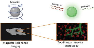
Multimodal imaging approach to track theranostic nanoparticle accumulation in glioblastoma with magnetic resonance imaging and intravital microscopy†
Theranostic nanoparticles (NPs) have been designed for simultaneous therapeutic and diagnostic purposes, thereby enabling personalized cancer therapy and in vivo drug tracking. However, studies thus far have focused on imaging NP tumor accumulation at the macroscopic level and correlating results with ex vivo histology. Limited evidence exists on whether in vivo NP tumor contrast enhancement on magnetic resonance imaging (MRI) correlates with in vivo NP tumor accumulation at the microscopic level. To address this gap, the purpose of our study was to correlate quantitative MRI estimates of NP accumulation with in vivo NP signal quantification as measured through two-photon intravital microscopy (IVM) in an orthotopic murine glioblastoma multiforme model (GBM). To enable multimodal imaging, we designed dual-mode NPs, composed of a carbohydrate-coated magnetic core (Ferumoxytol) as an MRI contrast agent, and a conjugated fluorophore (FITC) for IVM detection. We administered these NPs with or without a conjugated vascular disrupting agent (VDA) to assess its effect on NP delivery to GBM. We correlated in vivo MRI contrast enhancement in tumors, quantified as T2 relaxation time, with IVM fluorescence spatial decay rate. Results demonstrated a significantly lower tumor T2 relaxation time and spatial decay rate in tumors targeted with VDA-conjugated NPs compared to unconjugated NPs. Postmortem histological analyses validated the in vivo observations. The presented multimodal imaging approach enabled a quantitative correlation between MRI contrast enhancement at the macroscopic level and NP accumulation in the tumor microenvironment. These studies lay the groundwork for the precise evaluation of the tumor targeting of theranostic NPs.
期刊介绍:
Nanoscale is a high-impact international journal, publishing high-quality research across nanoscience and nanotechnology. Nanoscale publishes a full mix of research articles on experimental and theoretical work, including reviews, communications, and full papers.Highly interdisciplinary, this journal appeals to scientists, researchers and professionals interested in nanoscience and nanotechnology, quantum materials and quantum technology, including the areas of physics, chemistry, biology, medicine, materials, energy/environment, information technology, detection science, healthcare and drug discovery, and electronics.
 求助内容:
求助内容: 应助结果提醒方式:


