{"title":"根尖乳头细胞对受大肠杆菌脂多糖刺激的牙周韧带成纤维细胞的免疫调节作用:一项体外研究。","authors":"Alexandre Guimarães Dos Santos, Karollyne Santos Spigariol, Letícia Martins Santos, Marinella Holzhausen, Carla Renata Sipert","doi":"10.1590/1678-7757-2024-0338","DOIUrl":null,"url":null,"abstract":"<p><strong>Background: </strong>The role of human Stem Cells from the Apical Papilla (SCAP) in tissue regeneration has been described, but their impact on modulating the apical inflammatory process by other surrounding cell populations, such as periodontal ligament fibroblasts (PLFs), is unclear. Therefore, we investigated the role of SCAP in the activation of PLFs in vitro.</p><p><strong>Methods: </strong>Primary SCAP culture was used to obtain conditioned media (CM). A primary human PLF culture was established and stimulated with increasing concentrations of Escherichia coli lipopolysaccharide (LPS) (0.01, 0.1, and 1 µg/mL). At the 24 h time-point, an MTT viability assay was performed, and interleukin (IL)-6 and chemokine (CC-motif) ligand 2 (CCL2) levels were quantified by enzyme-linked immunosorbent assay. Then, PLFs were stimulated with LPS in the presence of SCAP-CM (1:5 dilution) for cell viability assessment and cytokine detection. The following groups were tested: PLF activated with LPS at concentrations of 0.01 and 1 µg/mL with or without SCAP-CM; a group with PLF stimulated by SCAP-CM alone; and a control group (proliferation medium only). The experiments were conducted in triplicate and sextuplicate. Statistical analyses were performed using analysis of variance followed by Tukey's post-hoc test, with statistical significance established at 5% (p=0.05).</p><p><strong>Results: </strong>The MTT assay showed no cytotoxicity of LPS or SCAP-CM on PLFs (p>0.05). The production of CCL2 and IL-6 significantly increased in the presence of SCAP-CM regardless of the presence of LPS (p<0.0001).</p><p><strong>Conclusion: </strong>SCAP-CM significantly enhanced the release of proinflammatory cytokines by PLFs in vitro.</p>","PeriodicalId":15133,"journal":{"name":"Journal of Applied Oral Science","volume":"33 ","pages":"e20240338"},"PeriodicalIF":2.6000,"publicationDate":"2025-03-14","publicationTypes":"Journal Article","fieldsOfStudy":null,"isOpenAccess":false,"openAccessPdf":"https://www.ncbi.nlm.nih.gov/pmc/articles/PMC11869941/pdf/","citationCount":"0","resultStr":"{\"title\":\"Immunomodulatory effects of apical papilla cells on periodontal ligament fibroblasts stimulated with Escherichia coli lipopolysaccharide: an in vitro study.\",\"authors\":\"Alexandre Guimarães Dos Santos, Karollyne Santos Spigariol, Letícia Martins Santos, Marinella Holzhausen, Carla Renata Sipert\",\"doi\":\"10.1590/1678-7757-2024-0338\",\"DOIUrl\":null,\"url\":null,\"abstract\":\"<p><strong>Background: </strong>The role of human Stem Cells from the Apical Papilla (SCAP) in tissue regeneration has been described, but their impact on modulating the apical inflammatory process by other surrounding cell populations, such as periodontal ligament fibroblasts (PLFs), is unclear. Therefore, we investigated the role of SCAP in the activation of PLFs in vitro.</p><p><strong>Methods: </strong>Primary SCAP culture was used to obtain conditioned media (CM). A primary human PLF culture was established and stimulated with increasing concentrations of Escherichia coli lipopolysaccharide (LPS) (0.01, 0.1, and 1 µg/mL). At the 24 h time-point, an MTT viability assay was performed, and interleukin (IL)-6 and chemokine (CC-motif) ligand 2 (CCL2) levels were quantified by enzyme-linked immunosorbent assay. Then, PLFs were stimulated with LPS in the presence of SCAP-CM (1:5 dilution) for cell viability assessment and cytokine detection. The following groups were tested: PLF activated with LPS at concentrations of 0.01 and 1 µg/mL with or without SCAP-CM; a group with PLF stimulated by SCAP-CM alone; and a control group (proliferation medium only). The experiments were conducted in triplicate and sextuplicate. Statistical analyses were performed using analysis of variance followed by Tukey's post-hoc test, with statistical significance established at 5% (p=0.05).</p><p><strong>Results: </strong>The MTT assay showed no cytotoxicity of LPS or SCAP-CM on PLFs (p>0.05). The production of CCL2 and IL-6 significantly increased in the presence of SCAP-CM regardless of the presence of LPS (p<0.0001).</p><p><strong>Conclusion: </strong>SCAP-CM significantly enhanced the release of proinflammatory cytokines by PLFs in vitro.</p>\",\"PeriodicalId\":15133,\"journal\":{\"name\":\"Journal of Applied Oral Science\",\"volume\":\"33 \",\"pages\":\"e20240338\"},\"PeriodicalIF\":2.6000,\"publicationDate\":\"2025-03-14\",\"publicationTypes\":\"Journal Article\",\"fieldsOfStudy\":null,\"isOpenAccess\":false,\"openAccessPdf\":\"https://www.ncbi.nlm.nih.gov/pmc/articles/PMC11869941/pdf/\",\"citationCount\":\"0\",\"resultStr\":null,\"platform\":\"Semanticscholar\",\"paperid\":null,\"PeriodicalName\":\"Journal of Applied Oral Science\",\"FirstCategoryId\":\"3\",\"ListUrlMain\":\"https://doi.org/10.1590/1678-7757-2024-0338\",\"RegionNum\":3,\"RegionCategory\":\"医学\",\"ArticlePicture\":[],\"TitleCN\":null,\"AbstractTextCN\":null,\"PMCID\":null,\"EPubDate\":\"2025/1/1 0:00:00\",\"PubModel\":\"eCollection\",\"JCR\":\"Q2\",\"JCRName\":\"DENTISTRY, ORAL SURGERY & MEDICINE\",\"Score\":null,\"Total\":0}","platform":"Semanticscholar","paperid":null,"PeriodicalName":"Journal of Applied Oral Science","FirstCategoryId":"3","ListUrlMain":"https://doi.org/10.1590/1678-7757-2024-0338","RegionNum":3,"RegionCategory":"医学","ArticlePicture":[],"TitleCN":null,"AbstractTextCN":null,"PMCID":null,"EPubDate":"2025/1/1 0:00:00","PubModel":"eCollection","JCR":"Q2","JCRName":"DENTISTRY, ORAL SURGERY & MEDICINE","Score":null,"Total":0}
Immunomodulatory effects of apical papilla cells on periodontal ligament fibroblasts stimulated with Escherichia coli lipopolysaccharide: an in vitro study.
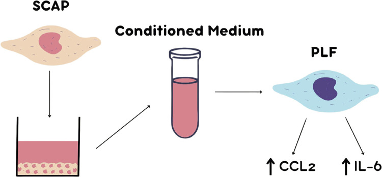
Background: The role of human Stem Cells from the Apical Papilla (SCAP) in tissue regeneration has been described, but their impact on modulating the apical inflammatory process by other surrounding cell populations, such as periodontal ligament fibroblasts (PLFs), is unclear. Therefore, we investigated the role of SCAP in the activation of PLFs in vitro.
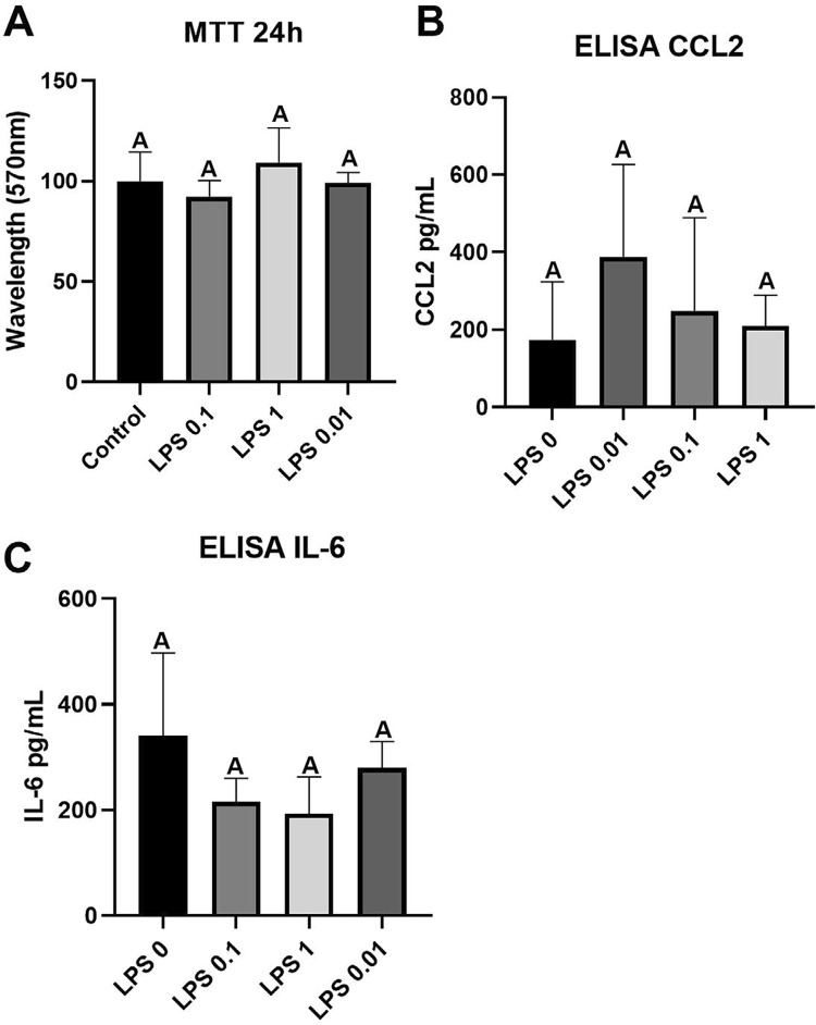
Methods: Primary SCAP culture was used to obtain conditioned media (CM). A primary human PLF culture was established and stimulated with increasing concentrations of Escherichia coli lipopolysaccharide (LPS) (0.01, 0.1, and 1 µg/mL). At the 24 h time-point, an MTT viability assay was performed, and interleukin (IL)-6 and chemokine (CC-motif) ligand 2 (CCL2) levels were quantified by enzyme-linked immunosorbent assay. Then, PLFs were stimulated with LPS in the presence of SCAP-CM (1:5 dilution) for cell viability assessment and cytokine detection. The following groups were tested: PLF activated with LPS at concentrations of 0.01 and 1 µg/mL with or without SCAP-CM; a group with PLF stimulated by SCAP-CM alone; and a control group (proliferation medium only). The experiments were conducted in triplicate and sextuplicate. Statistical analyses were performed using analysis of variance followed by Tukey's post-hoc test, with statistical significance established at 5% (p=0.05).
Results: The MTT assay showed no cytotoxicity of LPS or SCAP-CM on PLFs (p>0.05). The production of CCL2 and IL-6 significantly increased in the presence of SCAP-CM regardless of the presence of LPS (p<0.0001).
Conclusion: SCAP-CM significantly enhanced the release of proinflammatory cytokines by PLFs in vitro.
期刊介绍:
The Journal of Applied Oral Science is committed in publishing the scientific and technologic advances achieved by the dental community, according to the quality indicators and peer reviewed material, with the objective of assuring its acceptability at the local, regional, national and international levels. The primary goal of The Journal of Applied Oral Science is to publish the outcomes of original investigations as well as invited case reports and invited reviews in the field of Dentistry and related areas.
 求助内容:
求助内容: 应助结果提醒方式:


