Daniel H Lench, Aaron Embry, Niloufar Malakouti, Nathan DeTurk, Gonzalo J Revuelta
{"title":"步态冻结的自动性降低与皮质-小脑连通性升高有关。","authors":"Daniel H Lench, Aaron Embry, Niloufar Malakouti, Nathan DeTurk, Gonzalo J Revuelta","doi":"10.1007/s11682-025-00996-w","DOIUrl":null,"url":null,"abstract":"<p><p>Freezing of gait (FOG) in individuals with Parkinson's Disease is associated with a loss of gait automaticity. This loss of automaticity is demonstrated by worsening gait performance while dual tasking. Functional connectivity between the cerebellar vermis and cortex have previously been associated with spatiotemporal measures of gait in PD. The objective of this study was to determine whether this corticocerebellar connectivity is associated with gait automaticity as measured by dual task interference in PD FOG. 55 participants with PD were recruited (38 FOG, 17 non-FOG controls) to undergo a resting-state functional magnetic resonance imaging scan. Gait automaticity was quantified using spatiotemporal metrics from single and dual task time up and go trials. FOG participants demonstrated shorter step length and gait velocity compared to non-FOG PD controls. A trend toward greater dual task interference of step length in the FOG group was found. Using a seed-based connectivity approach we observed that FOG participants have greater vermis connectivity than non-FOG PD participants to several cortical regions including the superior parietal lobe, supplemental motor area, precentral gyrus and posterior cingulate (voxel threshold p < 0.01, cluster FWE corrected p < 0.05). Meanwhile, vermis connectivity to the occipital cortex was reduced in FOG participants relative to non-FOG controls. Dual task interference of step length among the FOG group correlated with the degree of vermis connectivity to the sensorimotor cortex and superior parietal cortex (voxel threshold p < 0.01, cluster FWE corrected p < 0.05). We conclude that increased corticocerebellar connectivity may be associated with loss of gait automaticity in individuals with PD FOG.</p>","PeriodicalId":9192,"journal":{"name":"Brain Imaging and Behavior","volume":" ","pages":"637-646"},"PeriodicalIF":2.4000,"publicationDate":"2025-06-01","publicationTypes":"Journal Article","fieldsOfStudy":null,"isOpenAccess":false,"openAccessPdf":"https://www.ncbi.nlm.nih.gov/pmc/articles/PMC12198313/pdf/","citationCount":"0","resultStr":"{\"title\":\"Reduced automaticity in freezing of gait is associated with elevated cortico-cerebellar connectivity.\",\"authors\":\"Daniel H Lench, Aaron Embry, Niloufar Malakouti, Nathan DeTurk, Gonzalo J Revuelta\",\"doi\":\"10.1007/s11682-025-00996-w\",\"DOIUrl\":null,\"url\":null,\"abstract\":\"<p><p>Freezing of gait (FOG) in individuals with Parkinson's Disease is associated with a loss of gait automaticity. This loss of automaticity is demonstrated by worsening gait performance while dual tasking. Functional connectivity between the cerebellar vermis and cortex have previously been associated with spatiotemporal measures of gait in PD. The objective of this study was to determine whether this corticocerebellar connectivity is associated with gait automaticity as measured by dual task interference in PD FOG. 55 participants with PD were recruited (38 FOG, 17 non-FOG controls) to undergo a resting-state functional magnetic resonance imaging scan. Gait automaticity was quantified using spatiotemporal metrics from single and dual task time up and go trials. FOG participants demonstrated shorter step length and gait velocity compared to non-FOG PD controls. A trend toward greater dual task interference of step length in the FOG group was found. Using a seed-based connectivity approach we observed that FOG participants have greater vermis connectivity than non-FOG PD participants to several cortical regions including the superior parietal lobe, supplemental motor area, precentral gyrus and posterior cingulate (voxel threshold p < 0.01, cluster FWE corrected p < 0.05). Meanwhile, vermis connectivity to the occipital cortex was reduced in FOG participants relative to non-FOG controls. Dual task interference of step length among the FOG group correlated with the degree of vermis connectivity to the sensorimotor cortex and superior parietal cortex (voxel threshold p < 0.01, cluster FWE corrected p < 0.05). We conclude that increased corticocerebellar connectivity may be associated with loss of gait automaticity in individuals with PD FOG.</p>\",\"PeriodicalId\":9192,\"journal\":{\"name\":\"Brain Imaging and Behavior\",\"volume\":\" \",\"pages\":\"637-646\"},\"PeriodicalIF\":2.4000,\"publicationDate\":\"2025-06-01\",\"publicationTypes\":\"Journal Article\",\"fieldsOfStudy\":null,\"isOpenAccess\":false,\"openAccessPdf\":\"https://www.ncbi.nlm.nih.gov/pmc/articles/PMC12198313/pdf/\",\"citationCount\":\"0\",\"resultStr\":null,\"platform\":\"Semanticscholar\",\"paperid\":null,\"PeriodicalName\":\"Brain Imaging and Behavior\",\"FirstCategoryId\":\"3\",\"ListUrlMain\":\"https://doi.org/10.1007/s11682-025-00996-w\",\"RegionNum\":3,\"RegionCategory\":\"医学\",\"ArticlePicture\":[],\"TitleCN\":null,\"AbstractTextCN\":null,\"PMCID\":null,\"EPubDate\":\"2025/3/18 0:00:00\",\"PubModel\":\"Epub\",\"JCR\":\"Q2\",\"JCRName\":\"NEUROIMAGING\",\"Score\":null,\"Total\":0}","platform":"Semanticscholar","paperid":null,"PeriodicalName":"Brain Imaging and Behavior","FirstCategoryId":"3","ListUrlMain":"https://doi.org/10.1007/s11682-025-00996-w","RegionNum":3,"RegionCategory":"医学","ArticlePicture":[],"TitleCN":null,"AbstractTextCN":null,"PMCID":null,"EPubDate":"2025/3/18 0:00:00","PubModel":"Epub","JCR":"Q2","JCRName":"NEUROIMAGING","Score":null,"Total":0}
Reduced automaticity in freezing of gait is associated with elevated cortico-cerebellar connectivity.
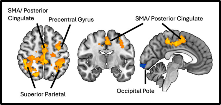
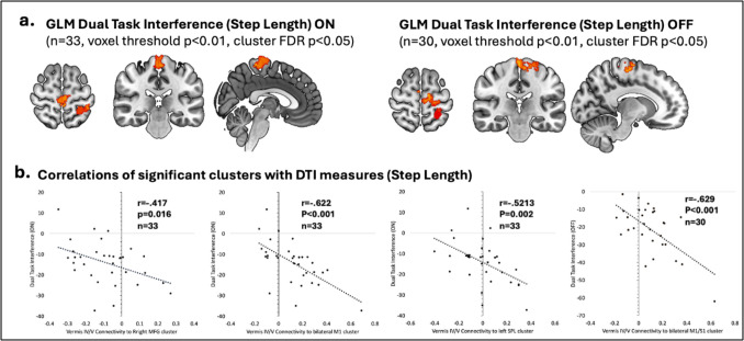
Freezing of gait (FOG) in individuals with Parkinson's Disease is associated with a loss of gait automaticity. This loss of automaticity is demonstrated by worsening gait performance while dual tasking. Functional connectivity between the cerebellar vermis and cortex have previously been associated with spatiotemporal measures of gait in PD. The objective of this study was to determine whether this corticocerebellar connectivity is associated with gait automaticity as measured by dual task interference in PD FOG. 55 participants with PD were recruited (38 FOG, 17 non-FOG controls) to undergo a resting-state functional magnetic resonance imaging scan. Gait automaticity was quantified using spatiotemporal metrics from single and dual task time up and go trials. FOG participants demonstrated shorter step length and gait velocity compared to non-FOG PD controls. A trend toward greater dual task interference of step length in the FOG group was found. Using a seed-based connectivity approach we observed that FOG participants have greater vermis connectivity than non-FOG PD participants to several cortical regions including the superior parietal lobe, supplemental motor area, precentral gyrus and posterior cingulate (voxel threshold p < 0.01, cluster FWE corrected p < 0.05). Meanwhile, vermis connectivity to the occipital cortex was reduced in FOG participants relative to non-FOG controls. Dual task interference of step length among the FOG group correlated with the degree of vermis connectivity to the sensorimotor cortex and superior parietal cortex (voxel threshold p < 0.01, cluster FWE corrected p < 0.05). We conclude that increased corticocerebellar connectivity may be associated with loss of gait automaticity in individuals with PD FOG.
期刊介绍:
Brain Imaging and Behavior is a bi-monthly, peer-reviewed journal, that publishes clinically relevant research using neuroimaging approaches to enhance our understanding of disorders of higher brain function. The journal is targeted at clinicians and researchers in fields concerned with human brain-behavior relationships, such as neuropsychology, psychiatry, neurology, neurosurgery, rehabilitation, and cognitive neuroscience.

 求助内容:
求助内容: 应助结果提醒方式:


