{"title":"一例青少年最佳黄斑营养不良伴脉络膜新生血管膜:1例报告及文献复习。","authors":"Yewande Olubunmi Babalola","doi":"10.4103/jwas.jwas_12_24","DOIUrl":null,"url":null,"abstract":"<p><p>An 11-year-old boy presented to the retina outpatient clinic with a -5-year history of poor vision in the left eye. The best corrected visual acuity at presentation was 6/5 and 6/36, respectively, in the right and left eyes. Ocular examination revealed normal anterior segments in both eyes. Binocular indirect ophthalmoscopy of the right eye revealed a pink disc with a cup disc ratio (CDR) of 0.3, normal vessels while the macula had a yellowish lesion with a scrambled egg appearance and surrounding dome-shaped subretinal fluid with a flat retina and no treatable peripheral retinal lesions. The left eye had a pink disc with CDR O.3, normal vessels with a hyperpigmented lesion at the macula surrounded by a small cuff of subretinal fluid with a flat retina and no treatable peripheral retinal lesions. Optical coherence tomography scan revealed subretinal fluid in both eyes with an active choroidal neovascular membrane in the left eye. He was advised on the need for left intravitreal anti-vascular endothelial growth factor injections.</p>","PeriodicalId":73993,"journal":{"name":"Journal of the West African College of Surgeons","volume":"15 2","pages":"240-242"},"PeriodicalIF":0.0000,"publicationDate":"2025-04-01","publicationTypes":"Journal Article","fieldsOfStudy":null,"isOpenAccess":false,"openAccessPdf":"https://www.ncbi.nlm.nih.gov/pmc/articles/PMC11908723/pdf/","citationCount":"0","resultStr":"{\"title\":\"Best Vitelliform Macular Dystrophy Presenting with Choroidal Neovascular Membrane in an Adolescent: A Case Report and a Review of the Literature.\",\"authors\":\"Yewande Olubunmi Babalola\",\"doi\":\"10.4103/jwas.jwas_12_24\",\"DOIUrl\":null,\"url\":null,\"abstract\":\"<p><p>An 11-year-old boy presented to the retina outpatient clinic with a -5-year history of poor vision in the left eye. The best corrected visual acuity at presentation was 6/5 and 6/36, respectively, in the right and left eyes. Ocular examination revealed normal anterior segments in both eyes. Binocular indirect ophthalmoscopy of the right eye revealed a pink disc with a cup disc ratio (CDR) of 0.3, normal vessels while the macula had a yellowish lesion with a scrambled egg appearance and surrounding dome-shaped subretinal fluid with a flat retina and no treatable peripheral retinal lesions. The left eye had a pink disc with CDR O.3, normal vessels with a hyperpigmented lesion at the macula surrounded by a small cuff of subretinal fluid with a flat retina and no treatable peripheral retinal lesions. Optical coherence tomography scan revealed subretinal fluid in both eyes with an active choroidal neovascular membrane in the left eye. He was advised on the need for left intravitreal anti-vascular endothelial growth factor injections.</p>\",\"PeriodicalId\":73993,\"journal\":{\"name\":\"Journal of the West African College of Surgeons\",\"volume\":\"15 2\",\"pages\":\"240-242\"},\"PeriodicalIF\":0.0000,\"publicationDate\":\"2025-04-01\",\"publicationTypes\":\"Journal Article\",\"fieldsOfStudy\":null,\"isOpenAccess\":false,\"openAccessPdf\":\"https://www.ncbi.nlm.nih.gov/pmc/articles/PMC11908723/pdf/\",\"citationCount\":\"0\",\"resultStr\":null,\"platform\":\"Semanticscholar\",\"paperid\":null,\"PeriodicalName\":\"Journal of the West African College of Surgeons\",\"FirstCategoryId\":\"1085\",\"ListUrlMain\":\"https://doi.org/10.4103/jwas.jwas_12_24\",\"RegionNum\":0,\"RegionCategory\":null,\"ArticlePicture\":[],\"TitleCN\":null,\"AbstractTextCN\":null,\"PMCID\":null,\"EPubDate\":\"2024/7/18 0:00:00\",\"PubModel\":\"Epub\",\"JCR\":\"\",\"JCRName\":\"\",\"Score\":null,\"Total\":0}","platform":"Semanticscholar","paperid":null,"PeriodicalName":"Journal of the West African College of Surgeons","FirstCategoryId":"1085","ListUrlMain":"https://doi.org/10.4103/jwas.jwas_12_24","RegionNum":0,"RegionCategory":null,"ArticlePicture":[],"TitleCN":null,"AbstractTextCN":null,"PMCID":null,"EPubDate":"2024/7/18 0:00:00","PubModel":"Epub","JCR":"","JCRName":"","Score":null,"Total":0}
Best Vitelliform Macular Dystrophy Presenting with Choroidal Neovascular Membrane in an Adolescent: A Case Report and a Review of the Literature.
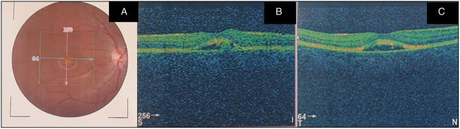
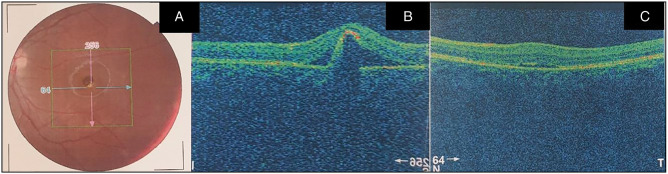
An 11-year-old boy presented to the retina outpatient clinic with a -5-year history of poor vision in the left eye. The best corrected visual acuity at presentation was 6/5 and 6/36, respectively, in the right and left eyes. Ocular examination revealed normal anterior segments in both eyes. Binocular indirect ophthalmoscopy of the right eye revealed a pink disc with a cup disc ratio (CDR) of 0.3, normal vessels while the macula had a yellowish lesion with a scrambled egg appearance and surrounding dome-shaped subretinal fluid with a flat retina and no treatable peripheral retinal lesions. The left eye had a pink disc with CDR O.3, normal vessels with a hyperpigmented lesion at the macula surrounded by a small cuff of subretinal fluid with a flat retina and no treatable peripheral retinal lesions. Optical coherence tomography scan revealed subretinal fluid in both eyes with an active choroidal neovascular membrane in the left eye. He was advised on the need for left intravitreal anti-vascular endothelial growth factor injections.

 求助内容:
求助内容: 应助结果提醒方式:


