{"title":"猪伪狂犬病毒感染江口萝卜仔猪脑组织转录组差异基因表达分析。","authors":"Wei Sun, Shan-Shan Liu, Yu Fan, Sheng-Qing Deng, Hua-Qi Zhang, Fengzhao Zhu, Shi-He Long, Tao-Hua Ren, Ling Bai","doi":"10.1177/20503121251326763","DOIUrl":null,"url":null,"abstract":"<p><strong>Background: </strong>The research focused on the effects of pseudorabies virus on gene expression in piglet brain tissue.</p><p><strong>Objectives: </strong>The goal was to understand the changes in gene expression in piglet brains due to pseudorabies virus infection.</p><p><strong>Design: </strong>The study used a comparative approach with infected and control piglet groups.</p><p><strong>Methods: </strong>Twelve 2-month-old piglets were divided into pseudorabies virus-infected and PBS-treated control groups, with brain tissue analyzed after 7 days.</p><p><strong>Results: </strong>Infected piglets showed increased oligodendrocyte counts and pseudorabies virus-positive signals. Transcriptomic analysis revealed 269 differentially expressed genes, with 149 up-regulated and 120 down-regulated. Gene ontology and Kyoto Encyclopedia of Genes and Genomes analyses indicated these genes are involved in signal transduction, transmembrane transport, apoptosis, and neuroactive ligand-receptor interaction. Quantitative fluorescent PCR validated these findings, particularly for genes related to neuroactive pathways, ferroptosis, and IL-17 signaling.</p><p><strong>Conclusion: </strong>The study provides valuable insights into the molecular alterations caused by pseudorabies virus in piglet brain tissue, enhancing our understanding of pseudorabies virus's pathogenic mechanisms.</p>","PeriodicalId":21398,"journal":{"name":"SAGE Open Medicine","volume":"13 ","pages":"20503121251326763"},"PeriodicalIF":2.1000,"publicationDate":"2025-03-14","publicationTypes":"Journal Article","fieldsOfStudy":null,"isOpenAccess":false,"openAccessPdf":"https://www.ncbi.nlm.nih.gov/pmc/articles/PMC11909670/pdf/","citationCount":"0","resultStr":"{\"title\":\"Analysis of differential gene expression in the brain tissue transcriptome of Jiangkou radish piglets infected with porcine pseudorabies virus.\",\"authors\":\"Wei Sun, Shan-Shan Liu, Yu Fan, Sheng-Qing Deng, Hua-Qi Zhang, Fengzhao Zhu, Shi-He Long, Tao-Hua Ren, Ling Bai\",\"doi\":\"10.1177/20503121251326763\",\"DOIUrl\":null,\"url\":null,\"abstract\":\"<p><strong>Background: </strong>The research focused on the effects of pseudorabies virus on gene expression in piglet brain tissue.</p><p><strong>Objectives: </strong>The goal was to understand the changes in gene expression in piglet brains due to pseudorabies virus infection.</p><p><strong>Design: </strong>The study used a comparative approach with infected and control piglet groups.</p><p><strong>Methods: </strong>Twelve 2-month-old piglets were divided into pseudorabies virus-infected and PBS-treated control groups, with brain tissue analyzed after 7 days.</p><p><strong>Results: </strong>Infected piglets showed increased oligodendrocyte counts and pseudorabies virus-positive signals. Transcriptomic analysis revealed 269 differentially expressed genes, with 149 up-regulated and 120 down-regulated. Gene ontology and Kyoto Encyclopedia of Genes and Genomes analyses indicated these genes are involved in signal transduction, transmembrane transport, apoptosis, and neuroactive ligand-receptor interaction. Quantitative fluorescent PCR validated these findings, particularly for genes related to neuroactive pathways, ferroptosis, and IL-17 signaling.</p><p><strong>Conclusion: </strong>The study provides valuable insights into the molecular alterations caused by pseudorabies virus in piglet brain tissue, enhancing our understanding of pseudorabies virus's pathogenic mechanisms.</p>\",\"PeriodicalId\":21398,\"journal\":{\"name\":\"SAGE Open Medicine\",\"volume\":\"13 \",\"pages\":\"20503121251326763\"},\"PeriodicalIF\":2.1000,\"publicationDate\":\"2025-03-14\",\"publicationTypes\":\"Journal Article\",\"fieldsOfStudy\":null,\"isOpenAccess\":false,\"openAccessPdf\":\"https://www.ncbi.nlm.nih.gov/pmc/articles/PMC11909670/pdf/\",\"citationCount\":\"0\",\"resultStr\":null,\"platform\":\"Semanticscholar\",\"paperid\":null,\"PeriodicalName\":\"SAGE Open Medicine\",\"FirstCategoryId\":\"1085\",\"ListUrlMain\":\"https://doi.org/10.1177/20503121251326763\",\"RegionNum\":0,\"RegionCategory\":null,\"ArticlePicture\":[],\"TitleCN\":null,\"AbstractTextCN\":null,\"PMCID\":null,\"EPubDate\":\"2025/1/1 0:00:00\",\"PubModel\":\"eCollection\",\"JCR\":\"Q2\",\"JCRName\":\"MEDICINE, GENERAL & INTERNAL\",\"Score\":null,\"Total\":0}","platform":"Semanticscholar","paperid":null,"PeriodicalName":"SAGE Open Medicine","FirstCategoryId":"1085","ListUrlMain":"https://doi.org/10.1177/20503121251326763","RegionNum":0,"RegionCategory":null,"ArticlePicture":[],"TitleCN":null,"AbstractTextCN":null,"PMCID":null,"EPubDate":"2025/1/1 0:00:00","PubModel":"eCollection","JCR":"Q2","JCRName":"MEDICINE, GENERAL & INTERNAL","Score":null,"Total":0}
Analysis of differential gene expression in the brain tissue transcriptome of Jiangkou radish piglets infected with porcine pseudorabies virus.
Background: The research focused on the effects of pseudorabies virus on gene expression in piglet brain tissue.
Objectives: The goal was to understand the changes in gene expression in piglet brains due to pseudorabies virus infection.
Design: The study used a comparative approach with infected and control piglet groups.
Methods: Twelve 2-month-old piglets were divided into pseudorabies virus-infected and PBS-treated control groups, with brain tissue analyzed after 7 days.
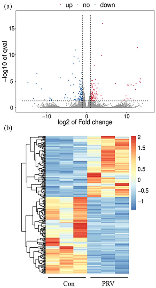
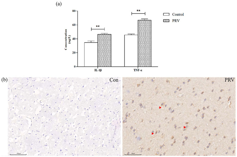
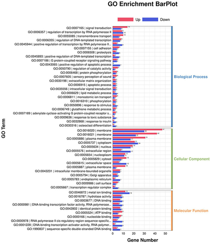
Results: Infected piglets showed increased oligodendrocyte counts and pseudorabies virus-positive signals. Transcriptomic analysis revealed 269 differentially expressed genes, with 149 up-regulated and 120 down-regulated. Gene ontology and Kyoto Encyclopedia of Genes and Genomes analyses indicated these genes are involved in signal transduction, transmembrane transport, apoptosis, and neuroactive ligand-receptor interaction. Quantitative fluorescent PCR validated these findings, particularly for genes related to neuroactive pathways, ferroptosis, and IL-17 signaling.
Conclusion: The study provides valuable insights into the molecular alterations caused by pseudorabies virus in piglet brain tissue, enhancing our understanding of pseudorabies virus's pathogenic mechanisms.

 求助内容:
求助内容: 应助结果提醒方式:


