Plern Sutra, Thananop Pothikamjorn, Sarah Lopez, Jaskirat Takhar, Mathinee Chongchareon, Jeremy Keenan, John A Gonzales
{"title":"前巩膜炎中巩膜紫色调的观察:前段光学相干断层扫描评价。","authors":"Plern Sutra, Thananop Pothikamjorn, Sarah Lopez, Jaskirat Takhar, Mathinee Chongchareon, Jeremy Keenan, John A Gonzales","doi":"10.1007/s00417-025-06788-8","DOIUrl":null,"url":null,"abstract":"<p><strong>Purpose: </strong>To determine the scleral thickness of inactive scleritis characterized by a violaceous hue (violaceous sclera) using anterior segment optical coherence tomography (AS-OCT).</p><p><strong>Methods: </strong>Retrospective observational case series of patients with inactive unilateral anterior scleritis featuring a violaceous hue. Mean scleral thickness was measured by AS-OCT in violaceous areas and compared with the same region in the contralateral unaffected eye. Measurements were performed by two masked graders.</p><p><strong>Results: </strong>Nine patients with median age of 52 ± 12.8 years were assessed. Eight patients were female. Rheumatoid arthritis and history of treated latent tuberculosis (33.3%) were the most common causes of anterior scleritis. Mean scleral thickness was 582.93 ± 124.03 µm and 648.59 ± 103.61 µm for violaceous sclera and the corresponding unaffected areas of the contralateral eye, respectively (mean difference = -65.65 µm, 95% CI: -143.73 to 12.42, p = 0.0885). The mean image contrast percentage of scleral hyperreflectivity as assessed by image conversion in an area of violaceous hue was 65.07 µm ± 6.49 µm compared to 42.70 µm ± 5.46 µm of unaffected areas (mean difference = 22.37 µm, 95% CI: 14.72 µm to 30.03 µm, p = 0.0001).</p><p><strong>Conclusion: </strong>Using AS-OCT, the thicknesses of violaceous sclerae were not significantly thinner than the contralateral unaffected areas, despite a mean difference of approximately 65 microns. The increased scleral hyperreflectivity observed in the violaceous sclera may suggest collagen remodeling in these areas. Such remodeling could play a role in the sclera reflecting violaceous hues while still preventing direct visualization of the underlying choroid.</p>","PeriodicalId":12795,"journal":{"name":"Graefe’s Archive for Clinical and Experimental Ophthalmology","volume":" ","pages":"1997-2004"},"PeriodicalIF":2.4000,"publicationDate":"2025-07-01","publicationTypes":"Journal Article","fieldsOfStudy":null,"isOpenAccess":false,"openAccessPdf":"https://www.ncbi.nlm.nih.gov/pmc/articles/PMC12373549/pdf/","citationCount":"0","resultStr":"{\"title\":\"Insights into scleral violaceous hue in anterior scleritis: anterior segment optical coherence tomography evaluation.\",\"authors\":\"Plern Sutra, Thananop Pothikamjorn, Sarah Lopez, Jaskirat Takhar, Mathinee Chongchareon, Jeremy Keenan, John A Gonzales\",\"doi\":\"10.1007/s00417-025-06788-8\",\"DOIUrl\":null,\"url\":null,\"abstract\":\"<p><strong>Purpose: </strong>To determine the scleral thickness of inactive scleritis characterized by a violaceous hue (violaceous sclera) using anterior segment optical coherence tomography (AS-OCT).</p><p><strong>Methods: </strong>Retrospective observational case series of patients with inactive unilateral anterior scleritis featuring a violaceous hue. Mean scleral thickness was measured by AS-OCT in violaceous areas and compared with the same region in the contralateral unaffected eye. Measurements were performed by two masked graders.</p><p><strong>Results: </strong>Nine patients with median age of 52 ± 12.8 years were assessed. Eight patients were female. Rheumatoid arthritis and history of treated latent tuberculosis (33.3%) were the most common causes of anterior scleritis. Mean scleral thickness was 582.93 ± 124.03 µm and 648.59 ± 103.61 µm for violaceous sclera and the corresponding unaffected areas of the contralateral eye, respectively (mean difference = -65.65 µm, 95% CI: -143.73 to 12.42, p = 0.0885). The mean image contrast percentage of scleral hyperreflectivity as assessed by image conversion in an area of violaceous hue was 65.07 µm ± 6.49 µm compared to 42.70 µm ± 5.46 µm of unaffected areas (mean difference = 22.37 µm, 95% CI: 14.72 µm to 30.03 µm, p = 0.0001).</p><p><strong>Conclusion: </strong>Using AS-OCT, the thicknesses of violaceous sclerae were not significantly thinner than the contralateral unaffected areas, despite a mean difference of approximately 65 microns. The increased scleral hyperreflectivity observed in the violaceous sclera may suggest collagen remodeling in these areas. Such remodeling could play a role in the sclera reflecting violaceous hues while still preventing direct visualization of the underlying choroid.</p>\",\"PeriodicalId\":12795,\"journal\":{\"name\":\"Graefe’s Archive for Clinical and Experimental Ophthalmology\",\"volume\":\" \",\"pages\":\"1997-2004\"},\"PeriodicalIF\":2.4000,\"publicationDate\":\"2025-07-01\",\"publicationTypes\":\"Journal Article\",\"fieldsOfStudy\":null,\"isOpenAccess\":false,\"openAccessPdf\":\"https://www.ncbi.nlm.nih.gov/pmc/articles/PMC12373549/pdf/\",\"citationCount\":\"0\",\"resultStr\":null,\"platform\":\"Semanticscholar\",\"paperid\":null,\"PeriodicalName\":\"Graefe’s Archive for Clinical and Experimental Ophthalmology\",\"FirstCategoryId\":\"3\",\"ListUrlMain\":\"https://doi.org/10.1007/s00417-025-06788-8\",\"RegionNum\":3,\"RegionCategory\":\"医学\",\"ArticlePicture\":[],\"TitleCN\":null,\"AbstractTextCN\":null,\"PMCID\":null,\"EPubDate\":\"2025/3/17 0:00:00\",\"PubModel\":\"Epub\",\"JCR\":\"Q2\",\"JCRName\":\"OPHTHALMOLOGY\",\"Score\":null,\"Total\":0}","platform":"Semanticscholar","paperid":null,"PeriodicalName":"Graefe’s Archive for Clinical and Experimental Ophthalmology","FirstCategoryId":"3","ListUrlMain":"https://doi.org/10.1007/s00417-025-06788-8","RegionNum":3,"RegionCategory":"医学","ArticlePicture":[],"TitleCN":null,"AbstractTextCN":null,"PMCID":null,"EPubDate":"2025/3/17 0:00:00","PubModel":"Epub","JCR":"Q2","JCRName":"OPHTHALMOLOGY","Score":null,"Total":0}
引用次数: 0
摘要
目的:利用前段光学相干断层扫描(AS-OCT)测定以紫色色相(紫色巩膜)为特征的非活动性巩膜炎的巩膜厚度。方法:回顾性观察单侧无活动性前巩膜炎呈紫色的病例系列。用AS-OCT测量对侧未受影响眼的平均巩膜厚度,并与对侧相同区域进行比较。测量由两名蒙面评分者完成。结果:共纳入9例患者,中位年龄52±12.8岁。女性8例。类风湿关节炎和潜伏性结核治疗史(33.3%)是前巩膜炎最常见的原因。对侧眼巩膜厚度平均值分别为582.93±124.03µm和648.59±103.61µm(平均差异= -65.65µm, 95% CI: -143.73 ~ 12.42, p = 0.0885)。通过图像转换评估,紫色区域巩膜高反射率的平均图像对比度为65.07µm±6.49µm,而未受影响区域为42.70µm±5.46µm(平均差异= 22.37µm, 95% CI: 14.72µm至30.03µm, p = 0.0001)。结论:在AS-OCT中,尽管平均相差约65微米,但侵犯巩膜厚度并未明显薄于对侧未受影响区域。紫巩膜高反射率的增加可能提示这些区域的胶原重塑。这种重塑可能在巩膜反射紫色的同时仍然阻止直接看到下面的脉络膜。
Insights into scleral violaceous hue in anterior scleritis: anterior segment optical coherence tomography evaluation.
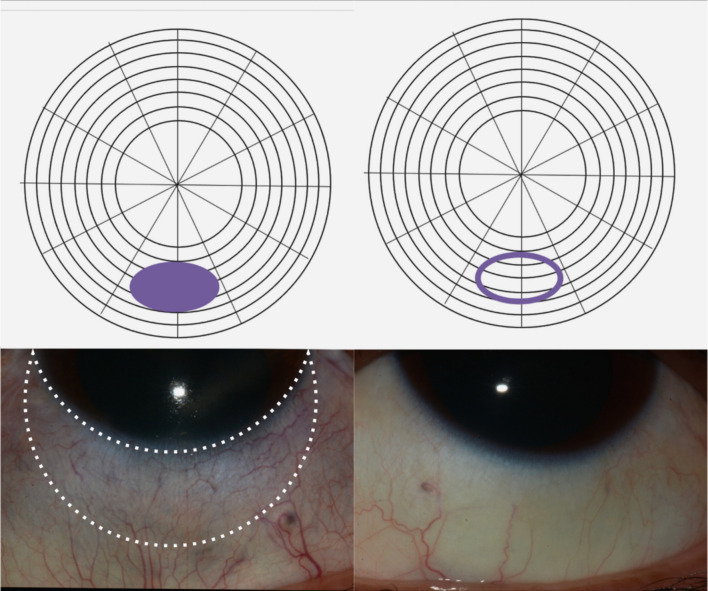
Purpose: To determine the scleral thickness of inactive scleritis characterized by a violaceous hue (violaceous sclera) using anterior segment optical coherence tomography (AS-OCT).
Methods: Retrospective observational case series of patients with inactive unilateral anterior scleritis featuring a violaceous hue. Mean scleral thickness was measured by AS-OCT in violaceous areas and compared with the same region in the contralateral unaffected eye. Measurements were performed by two masked graders.
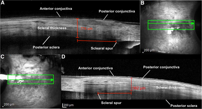
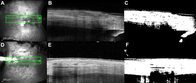
Results: Nine patients with median age of 52 ± 12.8 years were assessed. Eight patients were female. Rheumatoid arthritis and history of treated latent tuberculosis (33.3%) were the most common causes of anterior scleritis. Mean scleral thickness was 582.93 ± 124.03 µm and 648.59 ± 103.61 µm for violaceous sclera and the corresponding unaffected areas of the contralateral eye, respectively (mean difference = -65.65 µm, 95% CI: -143.73 to 12.42, p = 0.0885). The mean image contrast percentage of scleral hyperreflectivity as assessed by image conversion in an area of violaceous hue was 65.07 µm ± 6.49 µm compared to 42.70 µm ± 5.46 µm of unaffected areas (mean difference = 22.37 µm, 95% CI: 14.72 µm to 30.03 µm, p = 0.0001).
Conclusion: Using AS-OCT, the thicknesses of violaceous sclerae were not significantly thinner than the contralateral unaffected areas, despite a mean difference of approximately 65 microns. The increased scleral hyperreflectivity observed in the violaceous sclera may suggest collagen remodeling in these areas. Such remodeling could play a role in the sclera reflecting violaceous hues while still preventing direct visualization of the underlying choroid.
期刊介绍:
Graefe''s Archive for Clinical and Experimental Ophthalmology is a distinguished international journal that presents original clinical reports and clini-cally relevant experimental studies. Founded in 1854 by Albrecht von Graefe to serve as a source of useful clinical information and a stimulus for discussion, the journal has published articles by leading ophthalmologists and vision research scientists for more than a century. With peer review by an international Editorial Board and prompt English-language publication, Graefe''s Archive provides rapid dissemination of clinical and clinically related experimental information.

 求助内容:
求助内容: 应助结果提醒方式:


