Ramkailash Gujar, Giulia Gregori, Rosa Dolz-Marco, Alessio Muzi, Jay Chhablani, Daniela Fruttini, Lorenzo Mangoni, Clara Rizzo, Cesare Mariotti, Marco Lupidi
{"title":"\"制定标准\":黄斑 OCT 血管造影不同采集模式分析。","authors":"Ramkailash Gujar, Giulia Gregori, Rosa Dolz-Marco, Alessio Muzi, Jay Chhablani, Daniela Fruttini, Lorenzo Mangoni, Clara Rizzo, Cesare Mariotti, Marco Lupidi","doi":"10.1186/s40942-025-00653-w","DOIUrl":null,"url":null,"abstract":"<p><strong>Purpose: </strong>This study aimed to determine the optimal OCT angiography (OCT-A) scanning pattern using the SPECTRALIS HRA-OCT2 device to evaluate macular microvasculature perfusion in healthy subjects.</p><p><strong>Methods: </strong>Healthy subjects were imaged using the SPECTRALIS OCT-A Module (Heidelberg Engineering) with the following scanning protocols: 10ºX10º-512 ART 7 [P1], 10ºX10º-256 ART 5 [P2], 10ºX10º-512 ART 5 [P3], and 15ºX10º-256 ART 5 [P4], all centered on the macula. Vessel perfusion density (VPD) and vessel length density (VLD) of the superficial vascular complex (SVC) were calculated using ImageJ software to evaluate the differences between scanning patterns. Three additional 10ºx1º, ART 7 high-density images were also obtained using the in-built software (SP-X1701 Update 3, based on Heyex Software Version 1.9.215.0 H) in the macular area and the VPD and VLD for all the three10ºx1º pattern size images with the corresponding area of pattern 1 image[P1]. Two retinal specialists conducted a blind qualitative assessment of the foveal avascular zone and image quality.</p><p><strong>Results: </strong>Twenty eyes from 20 consecutive healthy patients were included in the study. The mean VPD for P1, P2, P3, and P4 were 35.60, 31.67, 31.18, and 31.16, respectively. Mean VLD for P1, P2, P3, and P4 were 7.54, 5.86, 6.74, and 4.40, respectively. Significant differences were found between P1 and the other patterns for both the VPD and VLD, but not between P2, P3, and P4. VPD and VLD for 10ºx1º high-density images were 33.20 and 4.61, respectively, with significant VLD differences compared to P1, but not for VPD. P1 scored the highest and P4 the lowest in the qualitative assessments.</p><p><strong>Conclusions: </strong>The 10ºX10º-512 ART 7 pattern showed statistically significant qualitative superiority and appeared optimal for blood flow detection with reduced noise in quantitative assessments.</p>","PeriodicalId":14289,"journal":{"name":"International Journal of Retina and Vitreous","volume":"11 1","pages":"28"},"PeriodicalIF":2.4000,"publicationDate":"2025-03-13","publicationTypes":"Journal Article","fieldsOfStudy":null,"isOpenAccess":false,"openAccessPdf":"https://www.ncbi.nlm.nih.gov/pmc/articles/PMC11907780/pdf/","citationCount":"0","resultStr":"{\"title\":\"\\\"Setting the standard\\\": an analysis of different acquisition patterns for macular OCT-angiography.\",\"authors\":\"Ramkailash Gujar, Giulia Gregori, Rosa Dolz-Marco, Alessio Muzi, Jay Chhablani, Daniela Fruttini, Lorenzo Mangoni, Clara Rizzo, Cesare Mariotti, Marco Lupidi\",\"doi\":\"10.1186/s40942-025-00653-w\",\"DOIUrl\":null,\"url\":null,\"abstract\":\"<p><strong>Purpose: </strong>This study aimed to determine the optimal OCT angiography (OCT-A) scanning pattern using the SPECTRALIS HRA-OCT2 device to evaluate macular microvasculature perfusion in healthy subjects.</p><p><strong>Methods: </strong>Healthy subjects were imaged using the SPECTRALIS OCT-A Module (Heidelberg Engineering) with the following scanning protocols: 10ºX10º-512 ART 7 [P1], 10ºX10º-256 ART 5 [P2], 10ºX10º-512 ART 5 [P3], and 15ºX10º-256 ART 5 [P4], all centered on the macula. Vessel perfusion density (VPD) and vessel length density (VLD) of the superficial vascular complex (SVC) were calculated using ImageJ software to evaluate the differences between scanning patterns. Three additional 10ºx1º, ART 7 high-density images were also obtained using the in-built software (SP-X1701 Update 3, based on Heyex Software Version 1.9.215.0 H) in the macular area and the VPD and VLD for all the three10ºx1º pattern size images with the corresponding area of pattern 1 image[P1]. Two retinal specialists conducted a blind qualitative assessment of the foveal avascular zone and image quality.</p><p><strong>Results: </strong>Twenty eyes from 20 consecutive healthy patients were included in the study. The mean VPD for P1, P2, P3, and P4 were 35.60, 31.67, 31.18, and 31.16, respectively. Mean VLD for P1, P2, P3, and P4 were 7.54, 5.86, 6.74, and 4.40, respectively. Significant differences were found between P1 and the other patterns for both the VPD and VLD, but not between P2, P3, and P4. VPD and VLD for 10ºx1º high-density images were 33.20 and 4.61, respectively, with significant VLD differences compared to P1, but not for VPD. P1 scored the highest and P4 the lowest in the qualitative assessments.</p><p><strong>Conclusions: </strong>The 10ºX10º-512 ART 7 pattern showed statistically significant qualitative superiority and appeared optimal for blood flow detection with reduced noise in quantitative assessments.</p>\",\"PeriodicalId\":14289,\"journal\":{\"name\":\"International Journal of Retina and Vitreous\",\"volume\":\"11 1\",\"pages\":\"28\"},\"PeriodicalIF\":2.4000,\"publicationDate\":\"2025-03-13\",\"publicationTypes\":\"Journal Article\",\"fieldsOfStudy\":null,\"isOpenAccess\":false,\"openAccessPdf\":\"https://www.ncbi.nlm.nih.gov/pmc/articles/PMC11907780/pdf/\",\"citationCount\":\"0\",\"resultStr\":null,\"platform\":\"Semanticscholar\",\"paperid\":null,\"PeriodicalName\":\"International Journal of Retina and Vitreous\",\"FirstCategoryId\":\"1085\",\"ListUrlMain\":\"https://doi.org/10.1186/s40942-025-00653-w\",\"RegionNum\":0,\"RegionCategory\":null,\"ArticlePicture\":[],\"TitleCN\":null,\"AbstractTextCN\":null,\"PMCID\":null,\"EPubDate\":\"\",\"PubModel\":\"\",\"JCR\":\"Q2\",\"JCRName\":\"OPHTHALMOLOGY\",\"Score\":null,\"Total\":0}","platform":"Semanticscholar","paperid":null,"PeriodicalName":"International Journal of Retina and Vitreous","FirstCategoryId":"1085","ListUrlMain":"https://doi.org/10.1186/s40942-025-00653-w","RegionNum":0,"RegionCategory":null,"ArticlePicture":[],"TitleCN":null,"AbstractTextCN":null,"PMCID":null,"EPubDate":"","PubModel":"","JCR":"Q2","JCRName":"OPHTHALMOLOGY","Score":null,"Total":0}
引用次数: 0
摘要
目的:本研究旨在利用SPECTRALIS hla - oct2设备确定最佳的OCT血管造影(OCT- a)扫描模式来评估健康受试者的黄斑微血管灌注。方法:健康受试者使用SPECTRALIS OCT-A模块(Heidelberg Engineering)进行扫描,扫描方案为:10ºX10º-512 ART 7 [P1], 10ºX10º-256 ART 5 [P2], 10ºX10º-512 ART 5 [P3]和15ºX10º-256 ART 5 [P4],均以黄斑为中心。采用ImageJ软件计算血管灌注密度(VPD)和血管长度密度(VLD),评价两种扫描方式的差异。使用内置软件(SP-X1701 Update 3,基于Heyex软件版本1.9.215.0 H)在黄斑区域获得另外3张10ºx1º、ART 7高密度图像,并获得所有3张10ºx1º模式尺寸图像与模式1图像对应区域的VPD和VLD [P1]。两位视网膜专家对中央凹无血管区和图像质量进行了盲定性评估。结果:20例连续健康患者的20只眼被纳入研究。P1、P2、P3、P4的平均VPD分别为35.60、31.67、31.18、31.16。P1、P2、P3和P4的平均VLD分别为7.54、5.86、6.74和4.40。VPD和VLD在P1和其他模式之间均有显著差异,但在P2、P3和P4之间无显著差异。10ºx1º高密度图像的VPD和VLD分别为33.20和4.61,VLD与P1有显著差异,VPD与P1无显著差异。在定性评价中,P1得分最高,P4得分最低。结论:10ºX10º-512 ART 7模式具有统计学上显著的定性优势,并且在定量评估中具有降低噪声的最佳血流检测方法。
"Setting the standard": an analysis of different acquisition patterns for macular OCT-angiography.
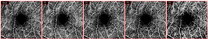
Purpose: This study aimed to determine the optimal OCT angiography (OCT-A) scanning pattern using the SPECTRALIS HRA-OCT2 device to evaluate macular microvasculature perfusion in healthy subjects.
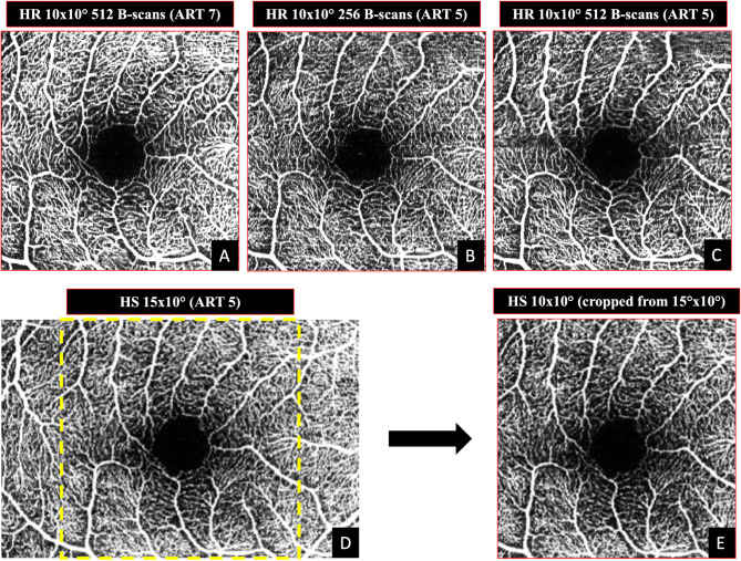
Methods: Healthy subjects were imaged using the SPECTRALIS OCT-A Module (Heidelberg Engineering) with the following scanning protocols: 10ºX10º-512 ART 7 [P1], 10ºX10º-256 ART 5 [P2], 10ºX10º-512 ART 5 [P3], and 15ºX10º-256 ART 5 [P4], all centered on the macula. Vessel perfusion density (VPD) and vessel length density (VLD) of the superficial vascular complex (SVC) were calculated using ImageJ software to evaluate the differences between scanning patterns. Three additional 10ºx1º, ART 7 high-density images were also obtained using the in-built software (SP-X1701 Update 3, based on Heyex Software Version 1.9.215.0 H) in the macular area and the VPD and VLD for all the three10ºx1º pattern size images with the corresponding area of pattern 1 image[P1]. Two retinal specialists conducted a blind qualitative assessment of the foveal avascular zone and image quality.
Results: Twenty eyes from 20 consecutive healthy patients were included in the study. The mean VPD for P1, P2, P3, and P4 were 35.60, 31.67, 31.18, and 31.16, respectively. Mean VLD for P1, P2, P3, and P4 were 7.54, 5.86, 6.74, and 4.40, respectively. Significant differences were found between P1 and the other patterns for both the VPD and VLD, but not between P2, P3, and P4. VPD and VLD for 10ºx1º high-density images were 33.20 and 4.61, respectively, with significant VLD differences compared to P1, but not for VPD. P1 scored the highest and P4 the lowest in the qualitative assessments.
Conclusions: The 10ºX10º-512 ART 7 pattern showed statistically significant qualitative superiority and appeared optimal for blood flow detection with reduced noise in quantitative assessments.
期刊介绍:
International Journal of Retina and Vitreous focuses on the ophthalmic subspecialty of vitreoretinal disorders. The journal presents original articles on new approaches to diagnosis, outcomes of clinical trials, innovations in pharmacological therapy and surgical techniques, as well as basic science advances that impact clinical practice. Topical areas include, but are not limited to: -Imaging of the retina, choroid and vitreous -Innovations in optical coherence tomography (OCT) -Small-gauge vitrectomy, retinal detachment, chromovitrectomy -Electroretinography (ERG), microperimetry, other functional tests -Intraocular tumors -Retinal pharmacotherapy & drug delivery -Diabetic retinopathy & other vascular diseases -Age-related macular degeneration (AMD) & other macular entities

 求助内容:
求助内容: 应助结果提醒方式:


