{"title":"第二版报告唾液腺细胞病理学的米兰系统:系统应用,结果和细胞组织学的相关性。","authors":"Magdalena Onyszczuk, Bogna Drozdzowska","doi":"10.4103/joc.joc_24_24","DOIUrl":null,"url":null,"abstract":"<p><strong>Background: </strong>Fine needle aspiration (FNA) cytology for salivary gland lesions is sensitive and specific for diagnosing and treating salivary gland pathologies. The objective of the Milan System for Reporting Salivary Gland Cytopathology (MSRSGC) is to organize the diagnostic information from the FNA into a uniform reporting terminology.</p><p><strong>Aims: </strong>The study was conducted retrospectively to reclassify previous diagnoses into the MSRSGC categories to determine the cytohistological concordance and assess the risk stratification by calculating the risk of malignancy (ROM) for different categories.</p><p><strong>Materials and methods: </strong>A total of 248 FNA cases of salivary gland lesions were analyzed and reclassified according to the second edition of the MSRSGC. The histological diagnosis was considered the gold standard. The ROM for each category was calculated based on 101 histopathologic follow-up cases.</p><p><strong>Results: </strong>Of the 248 patients, 1.2% were classified as nondiagnostic, 37.9% as nonneoplastic, 1.2% as atypia of undetermined significance (AUS), 52.8% as benign neoplasm, 0.4% as uncertain malignant potential (SUMP), 0.4% as suspicious of malignancy (SFM), and 6.1% as malignant neoplasm. Histopathological correlation was available in 101 cases. The ROM was 0% for nonneoplastic lesions and benign neoplasms, and 100% for AUS, SUMP, SFM, and malignant categories. The sensitivity, specificity, positive predictive value, and negative predictive value of FNA cytology in diagnosing salivary gland lesions using MSRSGC were found to be 76.5%, 100%, 100%, and 95.3%, respectively.</p><p><strong>Conclusion: </strong>The use of the MSRSGC helps in triaging patients with salivary gland lesions, increases the effectiveness of communication between clinicians and pathologists, and thus facilitates individualized patient management.</p>","PeriodicalId":50217,"journal":{"name":"Journal of Cytology","volume":"42 1","pages":"1-10"},"PeriodicalIF":1.0000,"publicationDate":"2024-01-01","publicationTypes":"Journal Article","fieldsOfStudy":null,"isOpenAccess":false,"openAccessPdf":"https://www.ncbi.nlm.nih.gov/pmc/articles/PMC11896119/pdf/","citationCount":"0","resultStr":"{\"title\":\"The Second Edition of the Milan System for Reporting Salivary Gland Cytopathology: System Application, Outcome, and Cytohistological Correlation.\",\"authors\":\"Magdalena Onyszczuk, Bogna Drozdzowska\",\"doi\":\"10.4103/joc.joc_24_24\",\"DOIUrl\":null,\"url\":null,\"abstract\":\"<p><strong>Background: </strong>Fine needle aspiration (FNA) cytology for salivary gland lesions is sensitive and specific for diagnosing and treating salivary gland pathologies. The objective of the Milan System for Reporting Salivary Gland Cytopathology (MSRSGC) is to organize the diagnostic information from the FNA into a uniform reporting terminology.</p><p><strong>Aims: </strong>The study was conducted retrospectively to reclassify previous diagnoses into the MSRSGC categories to determine the cytohistological concordance and assess the risk stratification by calculating the risk of malignancy (ROM) for different categories.</p><p><strong>Materials and methods: </strong>A total of 248 FNA cases of salivary gland lesions were analyzed and reclassified according to the second edition of the MSRSGC. The histological diagnosis was considered the gold standard. The ROM for each category was calculated based on 101 histopathologic follow-up cases.</p><p><strong>Results: </strong>Of the 248 patients, 1.2% were classified as nondiagnostic, 37.9% as nonneoplastic, 1.2% as atypia of undetermined significance (AUS), 52.8% as benign neoplasm, 0.4% as uncertain malignant potential (SUMP), 0.4% as suspicious of malignancy (SFM), and 6.1% as malignant neoplasm. Histopathological correlation was available in 101 cases. The ROM was 0% for nonneoplastic lesions and benign neoplasms, and 100% for AUS, SUMP, SFM, and malignant categories. The sensitivity, specificity, positive predictive value, and negative predictive value of FNA cytology in diagnosing salivary gland lesions using MSRSGC were found to be 76.5%, 100%, 100%, and 95.3%, respectively.</p><p><strong>Conclusion: </strong>The use of the MSRSGC helps in triaging patients with salivary gland lesions, increases the effectiveness of communication between clinicians and pathologists, and thus facilitates individualized patient management.</p>\",\"PeriodicalId\":50217,\"journal\":{\"name\":\"Journal of Cytology\",\"volume\":\"42 1\",\"pages\":\"1-10\"},\"PeriodicalIF\":1.0000,\"publicationDate\":\"2024-01-01\",\"publicationTypes\":\"Journal Article\",\"fieldsOfStudy\":null,\"isOpenAccess\":false,\"openAccessPdf\":\"https://www.ncbi.nlm.nih.gov/pmc/articles/PMC11896119/pdf/\",\"citationCount\":\"0\",\"resultStr\":null,\"platform\":\"Semanticscholar\",\"paperid\":null,\"PeriodicalName\":\"Journal of Cytology\",\"FirstCategoryId\":\"3\",\"ListUrlMain\":\"https://doi.org/10.4103/joc.joc_24_24\",\"RegionNum\":4,\"RegionCategory\":\"医学\",\"ArticlePicture\":[],\"TitleCN\":null,\"AbstractTextCN\":null,\"PMCID\":null,\"EPubDate\":\"2025/2/11 0:00:00\",\"PubModel\":\"Epub\",\"JCR\":\"Q4\",\"JCRName\":\"MEDICAL LABORATORY TECHNOLOGY\",\"Score\":null,\"Total\":0}","platform":"Semanticscholar","paperid":null,"PeriodicalName":"Journal of Cytology","FirstCategoryId":"3","ListUrlMain":"https://doi.org/10.4103/joc.joc_24_24","RegionNum":4,"RegionCategory":"医学","ArticlePicture":[],"TitleCN":null,"AbstractTextCN":null,"PMCID":null,"EPubDate":"2025/2/11 0:00:00","PubModel":"Epub","JCR":"Q4","JCRName":"MEDICAL LABORATORY TECHNOLOGY","Score":null,"Total":0}
The Second Edition of the Milan System for Reporting Salivary Gland Cytopathology: System Application, Outcome, and Cytohistological Correlation.
Background: Fine needle aspiration (FNA) cytology for salivary gland lesions is sensitive and specific for diagnosing and treating salivary gland pathologies. The objective of the Milan System for Reporting Salivary Gland Cytopathology (MSRSGC) is to organize the diagnostic information from the FNA into a uniform reporting terminology.
Aims: The study was conducted retrospectively to reclassify previous diagnoses into the MSRSGC categories to determine the cytohistological concordance and assess the risk stratification by calculating the risk of malignancy (ROM) for different categories.
Materials and methods: A total of 248 FNA cases of salivary gland lesions were analyzed and reclassified according to the second edition of the MSRSGC. The histological diagnosis was considered the gold standard. The ROM for each category was calculated based on 101 histopathologic follow-up cases.
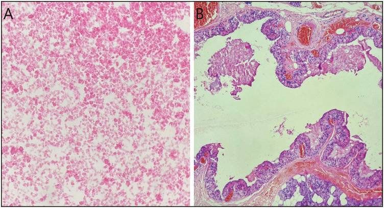
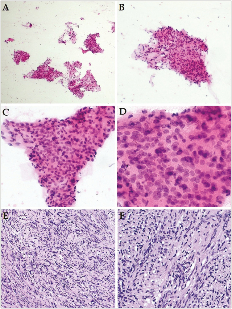
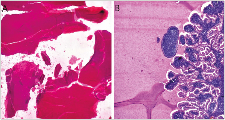
Results: Of the 248 patients, 1.2% were classified as nondiagnostic, 37.9% as nonneoplastic, 1.2% as atypia of undetermined significance (AUS), 52.8% as benign neoplasm, 0.4% as uncertain malignant potential (SUMP), 0.4% as suspicious of malignancy (SFM), and 6.1% as malignant neoplasm. Histopathological correlation was available in 101 cases. The ROM was 0% for nonneoplastic lesions and benign neoplasms, and 100% for AUS, SUMP, SFM, and malignant categories. The sensitivity, specificity, positive predictive value, and negative predictive value of FNA cytology in diagnosing salivary gland lesions using MSRSGC were found to be 76.5%, 100%, 100%, and 95.3%, respectively.
Conclusion: The use of the MSRSGC helps in triaging patients with salivary gland lesions, increases the effectiveness of communication between clinicians and pathologists, and thus facilitates individualized patient management.
期刊介绍:
The Journal of Cytology is the official Quarterly publication of the Indian Academy of Cytologists. It is in the 25th year of publication in the year 2008. The journal covers all aspects of diagnostic cytology, including fine needle aspiration cytology, gynecological and non-gynecological cytology. Articles on ancillary techniques, like cytochemistry, immunocytochemistry, electron microscopy, molecular cytopathology, as applied to cytological material are also welcome. The journal gives preference to clinically oriented studies over experimental and animal studies. The Journal would publish peer-reviewed original research papers, case reports, systematic reviews, meta-analysis, and debates.
 求助内容:
求助内容: 应助结果提醒方式:


