A Solderer, C Giuliani, D B Wiedemeier, R E Jung, P R Schmidlin
{"title":"组织水平种植体周围早期边缘种植体周围骨丢失:回顾性影像学评估。","authors":"A Solderer, C Giuliani, D B Wiedemeier, R E Jung, P R Schmidlin","doi":"10.1186/s40729-025-00613-x","DOIUrl":null,"url":null,"abstract":"<p><strong>Objectives: </strong>To retrospectively assess the potential impact of biological and host factors on radiographic bone loss following tissue-level implant placement and prosthetic rehabilitation.</p><p><strong>Methods: </strong>The University database was reviewed to identify patients treated with tissue-level implants between 2006 and 2020 at the University of Zurich, Switzerland. The study included patients who received screw-retained implant rehabilitations in the posterior area without simultaneous hard- or soft-tissue augmentations and had a follow-up period of at least 12 months. Radiographic measures of marginal bone loss and supracrestal tissue height were conducted using periapical x-rays at different time points. Additional factors analysed included age, gender, smoking status, history of periodontitis, jaw of treatment, type of reconstruction, and prosthetic emergence angle. Associations between marginal bone loss and potential explanatory variables were visualised and analysed. Elastic net regressions were applied to examine potential relationships with marginal bone loss.</p><p><strong>Results: </strong>A total of 1,479 patients were treated with tissue-level implants. After applying inclusion and exclusion criteria, 106 patients with 106 implants were included in the statistical evaluation after one year (T1, n = 106 implants), and 59 patients with 59 implants were evaluated after three years (T2, n = 59 implants). The mean marginal bone loss was 0.93 mm (SD 0.83) at T1 and 1.04 mm (SD 0.97) at T2. A strong correlation (Spearman) was found between mesial and distal bone loss. Smoking status and the jaw undergoing treatment were associated with bone loss. While these associations were observed in the univariate analysis, a more comprehensive multivariate analysis revealed that these variables had a limited effect on explaining radiographic bone loss.</p><p><strong>Conclusions: </strong>During the initial rehabilitation period in tissue-level implants in this cohort smoking status and jaw of treatment seemed to influence early peri-implant bone loss. Further, a strong correlation between mesial and distal MBL was observed. Additional research is required to determine factors contributing to early bone loss following implant-prosthetic rehabilitation.</p>","PeriodicalId":14076,"journal":{"name":"International Journal of Implant Dentistry","volume":"11 1","pages":"20"},"PeriodicalIF":4.0000,"publicationDate":"2025-03-12","publicationTypes":"Journal Article","fieldsOfStudy":null,"isOpenAccess":false,"openAccessPdf":"https://www.ncbi.nlm.nih.gov/pmc/articles/PMC11903984/pdf/","citationCount":"0","resultStr":"{\"title\":\"Early marginal peri-implant bone loss around tissue-level implants: a retrospective radiographic evaluation.\",\"authors\":\"A Solderer, C Giuliani, D B Wiedemeier, R E Jung, P R Schmidlin\",\"doi\":\"10.1186/s40729-025-00613-x\",\"DOIUrl\":null,\"url\":null,\"abstract\":\"<p><strong>Objectives: </strong>To retrospectively assess the potential impact of biological and host factors on radiographic bone loss following tissue-level implant placement and prosthetic rehabilitation.</p><p><strong>Methods: </strong>The University database was reviewed to identify patients treated with tissue-level implants between 2006 and 2020 at the University of Zurich, Switzerland. The study included patients who received screw-retained implant rehabilitations in the posterior area without simultaneous hard- or soft-tissue augmentations and had a follow-up period of at least 12 months. Radiographic measures of marginal bone loss and supracrestal tissue height were conducted using periapical x-rays at different time points. Additional factors analysed included age, gender, smoking status, history of periodontitis, jaw of treatment, type of reconstruction, and prosthetic emergence angle. Associations between marginal bone loss and potential explanatory variables were visualised and analysed. Elastic net regressions were applied to examine potential relationships with marginal bone loss.</p><p><strong>Results: </strong>A total of 1,479 patients were treated with tissue-level implants. After applying inclusion and exclusion criteria, 106 patients with 106 implants were included in the statistical evaluation after one year (T1, n = 106 implants), and 59 patients with 59 implants were evaluated after three years (T2, n = 59 implants). The mean marginal bone loss was 0.93 mm (SD 0.83) at T1 and 1.04 mm (SD 0.97) at T2. A strong correlation (Spearman) was found between mesial and distal bone loss. Smoking status and the jaw undergoing treatment were associated with bone loss. While these associations were observed in the univariate analysis, a more comprehensive multivariate analysis revealed that these variables had a limited effect on explaining radiographic bone loss.</p><p><strong>Conclusions: </strong>During the initial rehabilitation period in tissue-level implants in this cohort smoking status and jaw of treatment seemed to influence early peri-implant bone loss. Further, a strong correlation between mesial and distal MBL was observed. Additional research is required to determine factors contributing to early bone loss following implant-prosthetic rehabilitation.</p>\",\"PeriodicalId\":14076,\"journal\":{\"name\":\"International Journal of Implant Dentistry\",\"volume\":\"11 1\",\"pages\":\"20\"},\"PeriodicalIF\":4.0000,\"publicationDate\":\"2025-03-12\",\"publicationTypes\":\"Journal Article\",\"fieldsOfStudy\":null,\"isOpenAccess\":false,\"openAccessPdf\":\"https://www.ncbi.nlm.nih.gov/pmc/articles/PMC11903984/pdf/\",\"citationCount\":\"0\",\"resultStr\":null,\"platform\":\"Semanticscholar\",\"paperid\":null,\"PeriodicalName\":\"International Journal of Implant Dentistry\",\"FirstCategoryId\":\"3\",\"ListUrlMain\":\"https://doi.org/10.1186/s40729-025-00613-x\",\"RegionNum\":3,\"RegionCategory\":\"医学\",\"ArticlePicture\":[],\"TitleCN\":null,\"AbstractTextCN\":null,\"PMCID\":null,\"EPubDate\":\"\",\"PubModel\":\"\",\"JCR\":\"Q1\",\"JCRName\":\"DENTISTRY, ORAL SURGERY & MEDICINE\",\"Score\":null,\"Total\":0}","platform":"Semanticscholar","paperid":null,"PeriodicalName":"International Journal of Implant Dentistry","FirstCategoryId":"3","ListUrlMain":"https://doi.org/10.1186/s40729-025-00613-x","RegionNum":3,"RegionCategory":"医学","ArticlePicture":[],"TitleCN":null,"AbstractTextCN":null,"PMCID":null,"EPubDate":"","PubModel":"","JCR":"Q1","JCRName":"DENTISTRY, ORAL SURGERY & MEDICINE","Score":null,"Total":0}
引用次数: 0
摘要
目的:回顾性评估生物和宿主因素对组织水平种植体植入和假肢康复后放射学骨质流失的潜在影响。方法:回顾大学数据库,以确定2006年至2020年间在瑞士苏黎世大学接受组织级植入物治疗的患者。该研究纳入了在后侧区域接受螺钉保留种植体康复而没有同时进行硬组织或软组织增强的患者,随访期至少为12个月。在不同时间点使用根尖周x线进行边缘骨丢失和截骨上组织高度的x线测量。分析的其他因素包括年龄、性别、吸烟状况、牙周炎史、颌骨治疗、重建类型和假体出现角度。边缘骨质流失和潜在解释变量之间的关联被可视化和分析。弹性网回归应用于检查潜在的关系与边缘骨质流失。结果:1479例患者接受组织级种植体治疗。按照纳入和排除标准,106例106颗种植体在1年后(T1, n = 106颗)纳入统计评价,59例59颗种植体在3年后(T2, n = 59颗)进行统计评价。T1和T2分别为0.93 mm (SD 0.83)和1.04 mm (SD 0.97)。在近端和远端骨质流失之间有很强的相关性(Spearman)。吸烟状况和接受治疗的颌骨与骨质流失有关。虽然在单因素分析中观察到这些关联,但更全面的多因素分析显示,这些变量对解释x线骨质流失的影响有限。结论:在该队列中,在组织水平种植体的初始康复期,吸烟状态和颌骨治疗似乎影响早期种植体周围骨丢失。此外,观察到内侧和远端MBL之间有很强的相关性。需要进一步的研究来确定导致种植体修复后早期骨质流失的因素。
Early marginal peri-implant bone loss around tissue-level implants: a retrospective radiographic evaluation.
Objectives: To retrospectively assess the potential impact of biological and host factors on radiographic bone loss following tissue-level implant placement and prosthetic rehabilitation.
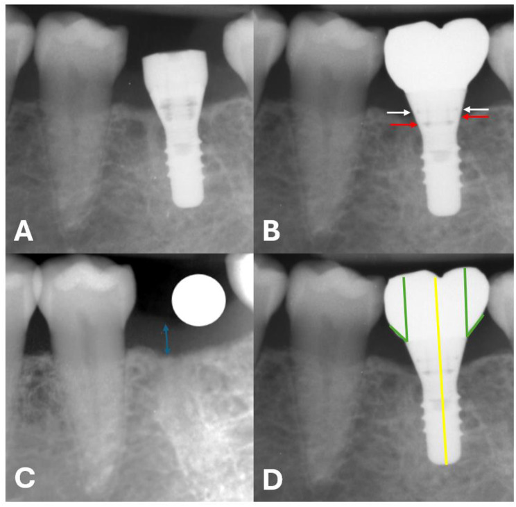
Methods: The University database was reviewed to identify patients treated with tissue-level implants between 2006 and 2020 at the University of Zurich, Switzerland. The study included patients who received screw-retained implant rehabilitations in the posterior area without simultaneous hard- or soft-tissue augmentations and had a follow-up period of at least 12 months. Radiographic measures of marginal bone loss and supracrestal tissue height were conducted using periapical x-rays at different time points. Additional factors analysed included age, gender, smoking status, history of periodontitis, jaw of treatment, type of reconstruction, and prosthetic emergence angle. Associations between marginal bone loss and potential explanatory variables were visualised and analysed. Elastic net regressions were applied to examine potential relationships with marginal bone loss.
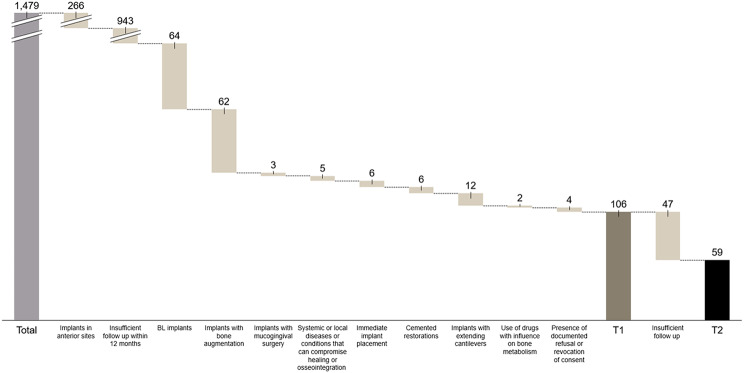
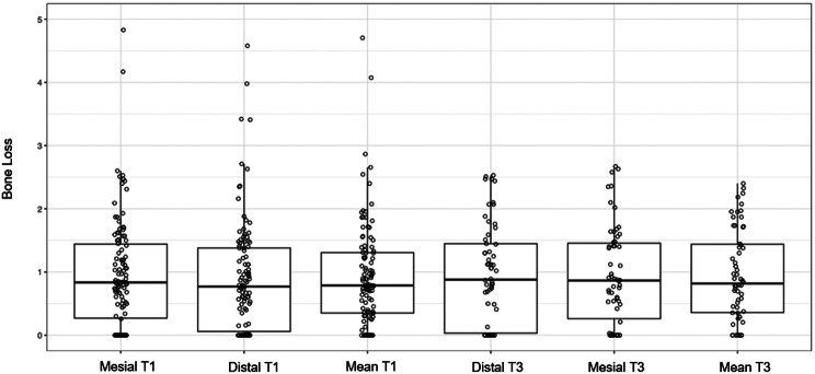
Results: A total of 1,479 patients were treated with tissue-level implants. After applying inclusion and exclusion criteria, 106 patients with 106 implants were included in the statistical evaluation after one year (T1, n = 106 implants), and 59 patients with 59 implants were evaluated after three years (T2, n = 59 implants). The mean marginal bone loss was 0.93 mm (SD 0.83) at T1 and 1.04 mm (SD 0.97) at T2. A strong correlation (Spearman) was found between mesial and distal bone loss. Smoking status and the jaw undergoing treatment were associated with bone loss. While these associations were observed in the univariate analysis, a more comprehensive multivariate analysis revealed that these variables had a limited effect on explaining radiographic bone loss.
Conclusions: During the initial rehabilitation period in tissue-level implants in this cohort smoking status and jaw of treatment seemed to influence early peri-implant bone loss. Further, a strong correlation between mesial and distal MBL was observed. Additional research is required to determine factors contributing to early bone loss following implant-prosthetic rehabilitation.
期刊介绍:
The International Journal of Implant Dentistry is a peer-reviewed open access journal published under the SpringerOpen brand. The journal is dedicated to promoting the exchange and discussion of all research areas relevant to implant dentistry in the form of systematic literature or invited reviews, prospective and retrospective clinical studies, clinical case reports, basic laboratory and animal research, and articles on material research and engineering.
 求助内容:
求助内容: 应助结果提醒方式:


