{"title":"抗牙龈卟啉单胞菌FimA型多克隆抗体在感染细胞中的内化和共定位","authors":"Napaporn Apiratmateekul, Kusuma Jamdee, Chantarapim Pinnork, Nawarat Khumyat, Niratcha Chaisomboon, Jantipa Jobsri","doi":"10.1055/s-0044-1801302","DOIUrl":null,"url":null,"abstract":"<p><p>The aim of this work was to investigate the effect of a rabbit polyclonal antibody specific to <i>Porphyromonas gingivalis</i> FimA type I (FimI) protein internalized into <i>P. gingivalis</i> infected cells.Rabbits were immunized with <i>P. gingivalis</i> FimI protein and the serum was collected for immunoglobulin (Ig) purification. For visualization of the antibody inside the cells, it was labeled with Cy3 dye. Live <i>P. gingivalis</i> was labeled with PKH67 dye. Rabbit anti-FimI Ig-Cy3 was internalized into H357 cells infected with <i>P. gingivalis</i>-PKH67 by electroporation or coincubation. Location of the Ig or <i>P. gingivalis</i> was observed under fluorescence microscope or confocal microscope. Percentage of <i>P. gingivalis</i>-PKH67 infected cells was analyzed by flow cytometry.Normality of data distribution was tested by Shapiro-Wilk test. The data failed normality test and were further analyzed by Kolmogorov-Smirnov test.Rabbit anti-<i>P. gingivalis</i> FimI Ig-Cy3 and <i>P. gingivalis</i>-PKH67 were both located next to the nucleus. The rabbit anti-FimI Ig-Cy3 was able to enter H357 cells after the cells were cultured in the medium containing the labeled Ig for 16 hours. The location of the Ig was near the nucleus as found in cells electroporated with the Ig-Cy3. The percentage of <i>P. gingivalis</i>-PKH67 infected cells seemed to be decreased after the infected cells internalized anti-FimI Ig by electroporation. However, it was not statistically significance.Rabbit anti-<i>P. gingivalis</i> FimI Ig and <i>P. gingivalis</i> was colocalized near the nucleus. And the rabbit anti-FimI Ig was able to enter H357 cells by coincubation method.</p>","PeriodicalId":12028,"journal":{"name":"European Journal of Dentistry","volume":" ","pages":"1078-1083"},"PeriodicalIF":2.1000,"publicationDate":"2025-10-01","publicationTypes":"Journal Article","fieldsOfStudy":null,"isOpenAccess":false,"openAccessPdf":"https://www.ncbi.nlm.nih.gov/pmc/articles/PMC12494417/pdf/","citationCount":"0","resultStr":"{\"title\":\"Internalization and Colocalization of a Polyclonal Antibody Against Porphyromonas gingivalis FimA type I in Infected Cells.\",\"authors\":\"Napaporn Apiratmateekul, Kusuma Jamdee, Chantarapim Pinnork, Nawarat Khumyat, Niratcha Chaisomboon, Jantipa Jobsri\",\"doi\":\"10.1055/s-0044-1801302\",\"DOIUrl\":null,\"url\":null,\"abstract\":\"<p><p>The aim of this work was to investigate the effect of a rabbit polyclonal antibody specific to <i>Porphyromonas gingivalis</i> FimA type I (FimI) protein internalized into <i>P. gingivalis</i> infected cells.Rabbits were immunized with <i>P. gingivalis</i> FimI protein and the serum was collected for immunoglobulin (Ig) purification. For visualization of the antibody inside the cells, it was labeled with Cy3 dye. Live <i>P. gingivalis</i> was labeled with PKH67 dye. Rabbit anti-FimI Ig-Cy3 was internalized into H357 cells infected with <i>P. gingivalis</i>-PKH67 by electroporation or coincubation. Location of the Ig or <i>P. gingivalis</i> was observed under fluorescence microscope or confocal microscope. Percentage of <i>P. gingivalis</i>-PKH67 infected cells was analyzed by flow cytometry.Normality of data distribution was tested by Shapiro-Wilk test. The data failed normality test and were further analyzed by Kolmogorov-Smirnov test.Rabbit anti-<i>P. gingivalis</i> FimI Ig-Cy3 and <i>P. gingivalis</i>-PKH67 were both located next to the nucleus. The rabbit anti-FimI Ig-Cy3 was able to enter H357 cells after the cells were cultured in the medium containing the labeled Ig for 16 hours. The location of the Ig was near the nucleus as found in cells electroporated with the Ig-Cy3. The percentage of <i>P. gingivalis</i>-PKH67 infected cells seemed to be decreased after the infected cells internalized anti-FimI Ig by electroporation. However, it was not statistically significance.Rabbit anti-<i>P. gingivalis</i> FimI Ig and <i>P. gingivalis</i> was colocalized near the nucleus. And the rabbit anti-FimI Ig was able to enter H357 cells by coincubation method.</p>\",\"PeriodicalId\":12028,\"journal\":{\"name\":\"European Journal of Dentistry\",\"volume\":\" \",\"pages\":\"1078-1083\"},\"PeriodicalIF\":2.1000,\"publicationDate\":\"2025-10-01\",\"publicationTypes\":\"Journal Article\",\"fieldsOfStudy\":null,\"isOpenAccess\":false,\"openAccessPdf\":\"https://www.ncbi.nlm.nih.gov/pmc/articles/PMC12494417/pdf/\",\"citationCount\":\"0\",\"resultStr\":null,\"platform\":\"Semanticscholar\",\"paperid\":null,\"PeriodicalName\":\"European Journal of Dentistry\",\"FirstCategoryId\":\"1085\",\"ListUrlMain\":\"https://doi.org/10.1055/s-0044-1801302\",\"RegionNum\":0,\"RegionCategory\":null,\"ArticlePicture\":[],\"TitleCN\":null,\"AbstractTextCN\":null,\"PMCID\":null,\"EPubDate\":\"2025/3/12 0:00:00\",\"PubModel\":\"Epub\",\"JCR\":\"Q1\",\"JCRName\":\"Dentistry\",\"Score\":null,\"Total\":0}","platform":"Semanticscholar","paperid":null,"PeriodicalName":"European Journal of Dentistry","FirstCategoryId":"1085","ListUrlMain":"https://doi.org/10.1055/s-0044-1801302","RegionNum":0,"RegionCategory":null,"ArticlePicture":[],"TitleCN":null,"AbstractTextCN":null,"PMCID":null,"EPubDate":"2025/3/12 0:00:00","PubModel":"Epub","JCR":"Q1","JCRName":"Dentistry","Score":null,"Total":0}
Internalization and Colocalization of a Polyclonal Antibody Against Porphyromonas gingivalis FimA type I in Infected Cells.
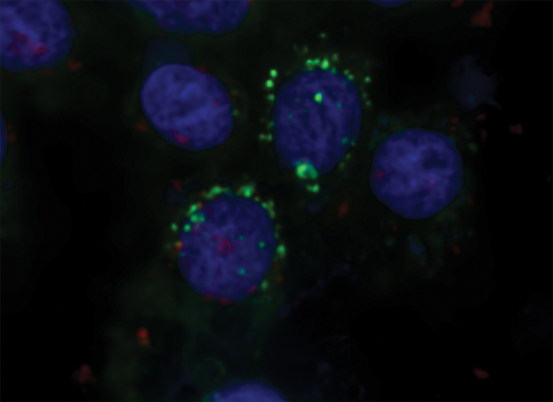
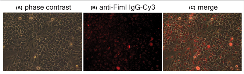
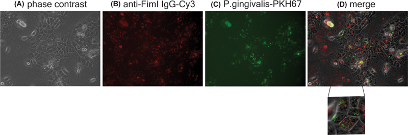
The aim of this work was to investigate the effect of a rabbit polyclonal antibody specific to Porphyromonas gingivalis FimA type I (FimI) protein internalized into P. gingivalis infected cells.Rabbits were immunized with P. gingivalis FimI protein and the serum was collected for immunoglobulin (Ig) purification. For visualization of the antibody inside the cells, it was labeled with Cy3 dye. Live P. gingivalis was labeled with PKH67 dye. Rabbit anti-FimI Ig-Cy3 was internalized into H357 cells infected with P. gingivalis-PKH67 by electroporation or coincubation. Location of the Ig or P. gingivalis was observed under fluorescence microscope or confocal microscope. Percentage of P. gingivalis-PKH67 infected cells was analyzed by flow cytometry.Normality of data distribution was tested by Shapiro-Wilk test. The data failed normality test and were further analyzed by Kolmogorov-Smirnov test.Rabbit anti-P. gingivalis FimI Ig-Cy3 and P. gingivalis-PKH67 were both located next to the nucleus. The rabbit anti-FimI Ig-Cy3 was able to enter H357 cells after the cells were cultured in the medium containing the labeled Ig for 16 hours. The location of the Ig was near the nucleus as found in cells electroporated with the Ig-Cy3. The percentage of P. gingivalis-PKH67 infected cells seemed to be decreased after the infected cells internalized anti-FimI Ig by electroporation. However, it was not statistically significance.Rabbit anti-P. gingivalis FimI Ig and P. gingivalis was colocalized near the nucleus. And the rabbit anti-FimI Ig was able to enter H357 cells by coincubation method.
期刊介绍:
The European Journal of Dentistry is the official journal of the Dental Investigations Society, based in Turkey. It is a double-blinded peer-reviewed, Open Access, multi-disciplinary international journal addressing various aspects of dentistry. The journal''s board consists of eminent investigators in dentistry from across the globe and presents an ideal international composition. The journal encourages its authors to submit original investigations, reviews, and reports addressing various divisions of dentistry including oral pathology, prosthodontics, endodontics, orthodontics etc. It is available both online and in print.

 求助内容:
求助内容: 应助结果提醒方式:


