Seyyed Ali Hosseini, Ghasem Hajianfar, Brandon Hall, Stijn Servaes, Pedro Rosa-Neto, Pardis Ghafarian, Habib Zaidi, Mohammad Reza Ay
{"title":"鲁棒与非鲁棒放射学特征:使用幻影和临床研究寻求最佳机器学习模型。","authors":"Seyyed Ali Hosseini, Ghasem Hajianfar, Brandon Hall, Stijn Servaes, Pedro Rosa-Neto, Pardis Ghafarian, Habib Zaidi, Mohammad Reza Ay","doi":"10.1186/s40644-025-00857-1","DOIUrl":null,"url":null,"abstract":"<p><strong>Purpose: </strong>This study aimed to select robust features against lung motion in a phantom study and use them as input to feature selection algorithms and machine learning classifiers in a clinical study to predict the lymphovascular invasion (LVI) of non-small cell lung cancer (NSCLC). The results of robust features were also compared with conventional techniques without considering the robustness of radiomic features.</p><p><strong>Methods: </strong>An in-house developed lung phantom was developed with two 22mm lesion sizes based on a clinical study. A specific motor was built to simulate motion in two orthogonal directions. Lesions of both clinical and phantom studies were segmented using a Fuzzy C-means-based segmentation algorithm. After inducing motion and extracting 105 radiomic features in 4 feature sets, including shape, first-, second-, and higher-order statistics features from each region of interest (ROI) of the phantom image, statistical analyses were performed to select robust features against motion. Subsequently, these robust features and a total of 105 radiomic features were extracted from 126 clinical data. Various feature selection (FS) and multiple machine learning (ML) classifiers were implemented to predict the LVI of NSCLC, followed by comparing the results of predicting LVI using robust features with common conventional techniques not considering the robustness of radiomic features.</p><p><strong>Results: </strong>Our results demonstrated that selecting robust features as input to FS algorithms and ML classifiers surges the sensitivity, which has a gentle negative effect on the accuracy and the area under the curve (AUC) of predictions compared with commonly used methods in 12 of 15 outcomes. The top performance of the LVI prediction was achieved by the NB classifier and RFE FS without considering the robustness of radiomic features with 95% area under the curve of AUC, 67% accuracy, and 100% sensitivity. Moreover, the top performance of the LVI prediction using robust features belonged to the NB classifier and Boruta feature selection with 92% AUC, 86% accuracy, and 100% sensitivity.</p><p><strong>Conclusion: </strong>Robustness over various influential factors is critical and should be considered in a radiomic study. Selecting robust features is a solution to overcome the low reproducibility of radiomic features. Although setting robust features against motion in a phantom study has a minor negative impact on the accuracy and AUC of LVI prediction, it boosts the sensitivity of prediction to a large extent.</p>","PeriodicalId":9548,"journal":{"name":"Cancer Imaging","volume":"25 1","pages":"33"},"PeriodicalIF":3.5000,"publicationDate":"2025-03-12","publicationTypes":"Journal Article","fieldsOfStudy":null,"isOpenAccess":false,"openAccessPdf":"https://www.ncbi.nlm.nih.gov/pmc/articles/PMC11905451/pdf/","citationCount":"0","resultStr":"{\"title\":\"Robust vs. Non-robust radiomic features: the quest for optimal machine learning models using phantom and clinical studies.\",\"authors\":\"Seyyed Ali Hosseini, Ghasem Hajianfar, Brandon Hall, Stijn Servaes, Pedro Rosa-Neto, Pardis Ghafarian, Habib Zaidi, Mohammad Reza Ay\",\"doi\":\"10.1186/s40644-025-00857-1\",\"DOIUrl\":null,\"url\":null,\"abstract\":\"<p><strong>Purpose: </strong>This study aimed to select robust features against lung motion in a phantom study and use them as input to feature selection algorithms and machine learning classifiers in a clinical study to predict the lymphovascular invasion (LVI) of non-small cell lung cancer (NSCLC). The results of robust features were also compared with conventional techniques without considering the robustness of radiomic features.</p><p><strong>Methods: </strong>An in-house developed lung phantom was developed with two 22mm lesion sizes based on a clinical study. A specific motor was built to simulate motion in two orthogonal directions. Lesions of both clinical and phantom studies were segmented using a Fuzzy C-means-based segmentation algorithm. After inducing motion and extracting 105 radiomic features in 4 feature sets, including shape, first-, second-, and higher-order statistics features from each region of interest (ROI) of the phantom image, statistical analyses were performed to select robust features against motion. Subsequently, these robust features and a total of 105 radiomic features were extracted from 126 clinical data. Various feature selection (FS) and multiple machine learning (ML) classifiers were implemented to predict the LVI of NSCLC, followed by comparing the results of predicting LVI using robust features with common conventional techniques not considering the robustness of radiomic features.</p><p><strong>Results: </strong>Our results demonstrated that selecting robust features as input to FS algorithms and ML classifiers surges the sensitivity, which has a gentle negative effect on the accuracy and the area under the curve (AUC) of predictions compared with commonly used methods in 12 of 15 outcomes. The top performance of the LVI prediction was achieved by the NB classifier and RFE FS without considering the robustness of radiomic features with 95% area under the curve of AUC, 67% accuracy, and 100% sensitivity. Moreover, the top performance of the LVI prediction using robust features belonged to the NB classifier and Boruta feature selection with 92% AUC, 86% accuracy, and 100% sensitivity.</p><p><strong>Conclusion: </strong>Robustness over various influential factors is critical and should be considered in a radiomic study. Selecting robust features is a solution to overcome the low reproducibility of radiomic features. Although setting robust features against motion in a phantom study has a minor negative impact on the accuracy and AUC of LVI prediction, it boosts the sensitivity of prediction to a large extent.</p>\",\"PeriodicalId\":9548,\"journal\":{\"name\":\"Cancer Imaging\",\"volume\":\"25 1\",\"pages\":\"33\"},\"PeriodicalIF\":3.5000,\"publicationDate\":\"2025-03-12\",\"publicationTypes\":\"Journal Article\",\"fieldsOfStudy\":null,\"isOpenAccess\":false,\"openAccessPdf\":\"https://www.ncbi.nlm.nih.gov/pmc/articles/PMC11905451/pdf/\",\"citationCount\":\"0\",\"resultStr\":null,\"platform\":\"Semanticscholar\",\"paperid\":null,\"PeriodicalName\":\"Cancer Imaging\",\"FirstCategoryId\":\"3\",\"ListUrlMain\":\"https://doi.org/10.1186/s40644-025-00857-1\",\"RegionNum\":2,\"RegionCategory\":\"医学\",\"ArticlePicture\":[],\"TitleCN\":null,\"AbstractTextCN\":null,\"PMCID\":null,\"EPubDate\":\"\",\"PubModel\":\"\",\"JCR\":\"Q2\",\"JCRName\":\"ONCOLOGY\",\"Score\":null,\"Total\":0}","platform":"Semanticscholar","paperid":null,"PeriodicalName":"Cancer Imaging","FirstCategoryId":"3","ListUrlMain":"https://doi.org/10.1186/s40644-025-00857-1","RegionNum":2,"RegionCategory":"医学","ArticlePicture":[],"TitleCN":null,"AbstractTextCN":null,"PMCID":null,"EPubDate":"","PubModel":"","JCR":"Q2","JCRName":"ONCOLOGY","Score":null,"Total":0}
Robust vs. Non-robust radiomic features: the quest for optimal machine learning models using phantom and clinical studies.
Purpose: This study aimed to select robust features against lung motion in a phantom study and use them as input to feature selection algorithms and machine learning classifiers in a clinical study to predict the lymphovascular invasion (LVI) of non-small cell lung cancer (NSCLC). The results of robust features were also compared with conventional techniques without considering the robustness of radiomic features.
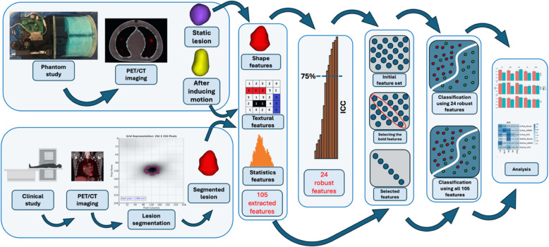
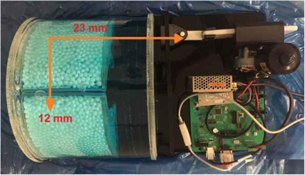
Methods: An in-house developed lung phantom was developed with two 22mm lesion sizes based on a clinical study. A specific motor was built to simulate motion in two orthogonal directions. Lesions of both clinical and phantom studies were segmented using a Fuzzy C-means-based segmentation algorithm. After inducing motion and extracting 105 radiomic features in 4 feature sets, including shape, first-, second-, and higher-order statistics features from each region of interest (ROI) of the phantom image, statistical analyses were performed to select robust features against motion. Subsequently, these robust features and a total of 105 radiomic features were extracted from 126 clinical data. Various feature selection (FS) and multiple machine learning (ML) classifiers were implemented to predict the LVI of NSCLC, followed by comparing the results of predicting LVI using robust features with common conventional techniques not considering the robustness of radiomic features.
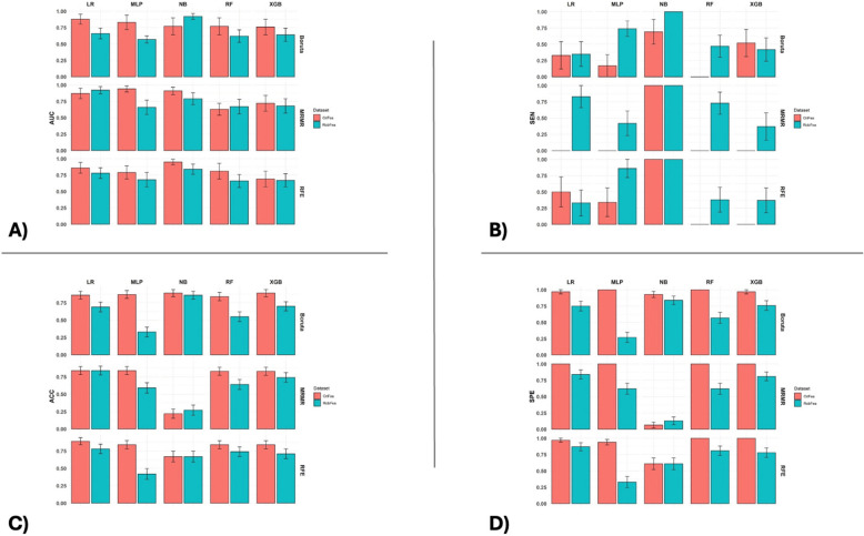
Results: Our results demonstrated that selecting robust features as input to FS algorithms and ML classifiers surges the sensitivity, which has a gentle negative effect on the accuracy and the area under the curve (AUC) of predictions compared with commonly used methods in 12 of 15 outcomes. The top performance of the LVI prediction was achieved by the NB classifier and RFE FS without considering the robustness of radiomic features with 95% area under the curve of AUC, 67% accuracy, and 100% sensitivity. Moreover, the top performance of the LVI prediction using robust features belonged to the NB classifier and Boruta feature selection with 92% AUC, 86% accuracy, and 100% sensitivity.
Conclusion: Robustness over various influential factors is critical and should be considered in a radiomic study. Selecting robust features is a solution to overcome the low reproducibility of radiomic features. Although setting robust features against motion in a phantom study has a minor negative impact on the accuracy and AUC of LVI prediction, it boosts the sensitivity of prediction to a large extent.
Cancer ImagingONCOLOGY-RADIOLOGY, NUCLEAR MEDICINE & MEDICAL IMAGING
CiteScore
7.00
自引率
0.00%
发文量
66
审稿时长
>12 weeks
期刊介绍:
Cancer Imaging is an open access, peer-reviewed journal publishing original articles, reviews and editorials written by expert international radiologists working in oncology.
The journal encompasses CT, MR, PET, ultrasound, radionuclide and multimodal imaging in all kinds of malignant tumours, plus new developments, techniques and innovations. Topics of interest include:
Breast Imaging
Chest
Complications of treatment
Ear, Nose & Throat
Gastrointestinal
Hepatobiliary & Pancreatic
Imaging biomarkers
Interventional
Lymphoma
Measurement of tumour response
Molecular functional imaging
Musculoskeletal
Neuro oncology
Nuclear Medicine
Paediatric.

 求助内容:
求助内容: 应助结果提醒方式:


