Lihuan Dai, Jinxue Yin, Xin Xin, Chun Yao, Yongfang Tang, Xiaohong Xia, Yuanlin Chen, Shuying Lai, Guoliang Lu, Jie Huang, Purong Zhang, Jiansheng Li, Xiangguang Chen, Xi Zhong
{"title":"基于计算机断层扫描放射组学预测胃癌程序性死亡配体1表达状态的可解释机器学习模型。","authors":"Lihuan Dai, Jinxue Yin, Xin Xin, Chun Yao, Yongfang Tang, Xiaohong Xia, Yuanlin Chen, Shuying Lai, Guoliang Lu, Jie Huang, Purong Zhang, Jiansheng Li, Xiangguang Chen, Xi Zhong","doi":"10.1186/s40644-025-00855-3","DOIUrl":null,"url":null,"abstract":"<p><strong>Background: </strong>Programmed death ligand 1 (PD-L1) expression status, closely related to immunotherapy outcomes, is a reliable biomarker for screening patients who may benefit from immunotherapy. Here, we developed and validated an interpretable machine learning (ML) model based on contrast-enhanced computed tomography (CECT) radiomics for preoperatively predicting PD-L1 expression status in patients with gastric cancer (GC).</p><p><strong>Methods: </strong>We retrospectively recruited 285 GC patients who underwent CECT and PD-L1 detection from two medical centers. A PD-L1 combined positive score (CPS) of ≥ 5 was considered to indicate a high PD-L1 expression status. Patients from center 1 were divided into training (n = 143) and validation sets (n = 62), and patients from center 2 were considered a test set (n = 80). Radiomics features were extracted from venous-phase CT images. After feature reduction and selection, 11 ML algorithms were employed to develop predictive models, and their performance in predicting PD-L1 expression status was evaluated using areas under receiver operating characteristic curves (AUCs). SHapley Additive exPlanations (SHAP) were used to interpret the optimal model and visualize the decision-making process for a single individual.</p><p><strong>Results: </strong>Nine features significantly associated with PD-L1 expression status were ultimately selected to construct the predictive model. The light gradient-boosting machine (LGBM) model demonstrated the best performance for PD-L1 high expression status prediction in the training, validation, and test sets, with AUCs of 0.841(95% CI: 0.773, 0.908), 0.834 (95% CI:0.729, 0.939), and 0.822 (95% CI: 0.718, 0.926), respectively. The SHAP summary and bar plots illustrated that a feature's value affected the feature's impact attributed to the model. The SHAP waterfall plots were used to visualize the decision-making process for a single individual.</p><p><strong>Conclusion: </strong>Our CT radiomics-based LGBM model may aid in preoperatively predicting PD-L1 expression status in GC patients, and the SHAP method may improve the interpretability of this model.</p>","PeriodicalId":9548,"journal":{"name":"Cancer Imaging","volume":"25 1","pages":"31"},"PeriodicalIF":3.5000,"publicationDate":"2025-03-12","publicationTypes":"Journal Article","fieldsOfStudy":null,"isOpenAccess":false,"openAccessPdf":"https://www.ncbi.nlm.nih.gov/pmc/articles/PMC11905525/pdf/","citationCount":"0","resultStr":"{\"title\":\"An interpretable machine learning model based on computed tomography radiomics for predicting programmed death ligand 1 expression status in gastric cancer.\",\"authors\":\"Lihuan Dai, Jinxue Yin, Xin Xin, Chun Yao, Yongfang Tang, Xiaohong Xia, Yuanlin Chen, Shuying Lai, Guoliang Lu, Jie Huang, Purong Zhang, Jiansheng Li, Xiangguang Chen, Xi Zhong\",\"doi\":\"10.1186/s40644-025-00855-3\",\"DOIUrl\":null,\"url\":null,\"abstract\":\"<p><strong>Background: </strong>Programmed death ligand 1 (PD-L1) expression status, closely related to immunotherapy outcomes, is a reliable biomarker for screening patients who may benefit from immunotherapy. Here, we developed and validated an interpretable machine learning (ML) model based on contrast-enhanced computed tomography (CECT) radiomics for preoperatively predicting PD-L1 expression status in patients with gastric cancer (GC).</p><p><strong>Methods: </strong>We retrospectively recruited 285 GC patients who underwent CECT and PD-L1 detection from two medical centers. A PD-L1 combined positive score (CPS) of ≥ 5 was considered to indicate a high PD-L1 expression status. Patients from center 1 were divided into training (n = 143) and validation sets (n = 62), and patients from center 2 were considered a test set (n = 80). Radiomics features were extracted from venous-phase CT images. After feature reduction and selection, 11 ML algorithms were employed to develop predictive models, and their performance in predicting PD-L1 expression status was evaluated using areas under receiver operating characteristic curves (AUCs). SHapley Additive exPlanations (SHAP) were used to interpret the optimal model and visualize the decision-making process for a single individual.</p><p><strong>Results: </strong>Nine features significantly associated with PD-L1 expression status were ultimately selected to construct the predictive model. The light gradient-boosting machine (LGBM) model demonstrated the best performance for PD-L1 high expression status prediction in the training, validation, and test sets, with AUCs of 0.841(95% CI: 0.773, 0.908), 0.834 (95% CI:0.729, 0.939), and 0.822 (95% CI: 0.718, 0.926), respectively. The SHAP summary and bar plots illustrated that a feature's value affected the feature's impact attributed to the model. The SHAP waterfall plots were used to visualize the decision-making process for a single individual.</p><p><strong>Conclusion: </strong>Our CT radiomics-based LGBM model may aid in preoperatively predicting PD-L1 expression status in GC patients, and the SHAP method may improve the interpretability of this model.</p>\",\"PeriodicalId\":9548,\"journal\":{\"name\":\"Cancer Imaging\",\"volume\":\"25 1\",\"pages\":\"31\"},\"PeriodicalIF\":3.5000,\"publicationDate\":\"2025-03-12\",\"publicationTypes\":\"Journal Article\",\"fieldsOfStudy\":null,\"isOpenAccess\":false,\"openAccessPdf\":\"https://www.ncbi.nlm.nih.gov/pmc/articles/PMC11905525/pdf/\",\"citationCount\":\"0\",\"resultStr\":null,\"platform\":\"Semanticscholar\",\"paperid\":null,\"PeriodicalName\":\"Cancer Imaging\",\"FirstCategoryId\":\"3\",\"ListUrlMain\":\"https://doi.org/10.1186/s40644-025-00855-3\",\"RegionNum\":2,\"RegionCategory\":\"医学\",\"ArticlePicture\":[],\"TitleCN\":null,\"AbstractTextCN\":null,\"PMCID\":null,\"EPubDate\":\"\",\"PubModel\":\"\",\"JCR\":\"Q2\",\"JCRName\":\"ONCOLOGY\",\"Score\":null,\"Total\":0}","platform":"Semanticscholar","paperid":null,"PeriodicalName":"Cancer Imaging","FirstCategoryId":"3","ListUrlMain":"https://doi.org/10.1186/s40644-025-00855-3","RegionNum":2,"RegionCategory":"医学","ArticlePicture":[],"TitleCN":null,"AbstractTextCN":null,"PMCID":null,"EPubDate":"","PubModel":"","JCR":"Q2","JCRName":"ONCOLOGY","Score":null,"Total":0}
An interpretable machine learning model based on computed tomography radiomics for predicting programmed death ligand 1 expression status in gastric cancer.
Background: Programmed death ligand 1 (PD-L1) expression status, closely related to immunotherapy outcomes, is a reliable biomarker for screening patients who may benefit from immunotherapy. Here, we developed and validated an interpretable machine learning (ML) model based on contrast-enhanced computed tomography (CECT) radiomics for preoperatively predicting PD-L1 expression status in patients with gastric cancer (GC).
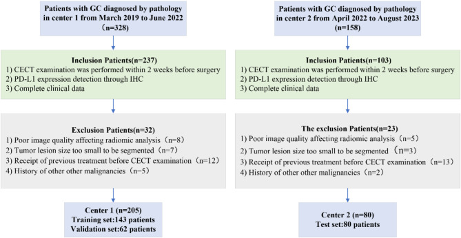
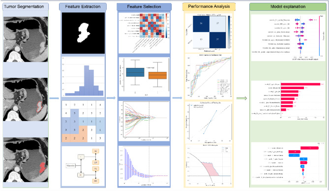
Methods: We retrospectively recruited 285 GC patients who underwent CECT and PD-L1 detection from two medical centers. A PD-L1 combined positive score (CPS) of ≥ 5 was considered to indicate a high PD-L1 expression status. Patients from center 1 were divided into training (n = 143) and validation sets (n = 62), and patients from center 2 were considered a test set (n = 80). Radiomics features were extracted from venous-phase CT images. After feature reduction and selection, 11 ML algorithms were employed to develop predictive models, and their performance in predicting PD-L1 expression status was evaluated using areas under receiver operating characteristic curves (AUCs). SHapley Additive exPlanations (SHAP) were used to interpret the optimal model and visualize the decision-making process for a single individual.
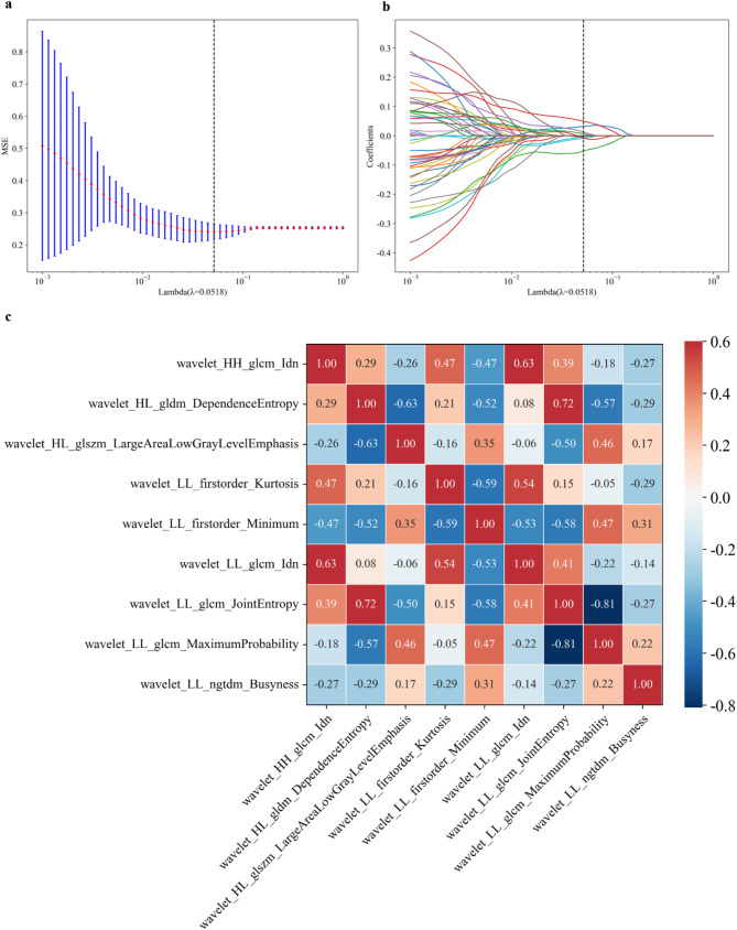
Results: Nine features significantly associated with PD-L1 expression status were ultimately selected to construct the predictive model. The light gradient-boosting machine (LGBM) model demonstrated the best performance for PD-L1 high expression status prediction in the training, validation, and test sets, with AUCs of 0.841(95% CI: 0.773, 0.908), 0.834 (95% CI:0.729, 0.939), and 0.822 (95% CI: 0.718, 0.926), respectively. The SHAP summary and bar plots illustrated that a feature's value affected the feature's impact attributed to the model. The SHAP waterfall plots were used to visualize the decision-making process for a single individual.
Conclusion: Our CT radiomics-based LGBM model may aid in preoperatively predicting PD-L1 expression status in GC patients, and the SHAP method may improve the interpretability of this model.
Cancer ImagingONCOLOGY-RADIOLOGY, NUCLEAR MEDICINE & MEDICAL IMAGING
CiteScore
7.00
自引率
0.00%
发文量
66
审稿时长
>12 weeks
期刊介绍:
Cancer Imaging is an open access, peer-reviewed journal publishing original articles, reviews and editorials written by expert international radiologists working in oncology.
The journal encompasses CT, MR, PET, ultrasound, radionuclide and multimodal imaging in all kinds of malignant tumours, plus new developments, techniques and innovations. Topics of interest include:
Breast Imaging
Chest
Complications of treatment
Ear, Nose & Throat
Gastrointestinal
Hepatobiliary & Pancreatic
Imaging biomarkers
Interventional
Lymphoma
Measurement of tumour response
Molecular functional imaging
Musculoskeletal
Neuro oncology
Nuclear Medicine
Paediatric.

 求助内容:
求助内容: 应助结果提醒方式:


