{"title":"韩国按解剖部位分布的术前影像深度预测脂肪瘤诊断准确性的回顾性分析。","authors":"Geon Hwi Kim, Jong Hun Lee","doi":"10.7181/acfs.2024.00535","DOIUrl":null,"url":null,"abstract":"<p><strong>Background: </strong>Lipomas are common benign connective-tissue tumors that usually present as slow-growing, painless, subcutaneous masses. Deeper variants, such as intramuscular, intermuscular, and submuscular lipomas, are larger and rarer. Accurate preoperative depth determination is crucial for planning appropriate surgical resection.</p><p><strong>Methods: </strong>We retrospectively reviewed 190 lipoma cases treated at a single medical center from January 2013 to August 2023. The accuracy of preoperative imaging techniques-ultrasonography (USG), computed tomography (CT), and magnetic resonance imaging (MRI)-in predicting lipoma depth was assessed.</p><p><strong>Results: </strong>USG, CT, and MRI showed accuracies of 72.5%, 56.5%, and 79.3%, respectively, with MRI showing the highest predictive accuracy. The trunk was the most common site for lipomas (49.5%), followed by the upper (20.5%) and lower extremities (13.2%). USG was more accurate for lipomas in the lower extremities and neck, whereas CT was less accurate for lipomas in the trunk.</p><p><strong>Conclusion: </strong>MRI is preferable for the preoperative depth assessment of lipomas, especially those located in the trunk. Accurate imaging is essential for guiding surgical planning and avoiding complications. Further studies with larger sample sizes are required to validate our findings.</p>","PeriodicalId":52238,"journal":{"name":"Archives of Craniofacial Surgery","volume":"26 1","pages":"13-18"},"PeriodicalIF":0.0000,"publicationDate":"2025-02-01","publicationTypes":"Journal Article","fieldsOfStudy":null,"isOpenAccess":false,"openAccessPdf":"https://www.ncbi.nlm.nih.gov/pmc/articles/PMC11917405/pdf/","citationCount":"0","resultStr":"{\"title\":\"The diagnostic accuracy of depth prediction for lipomas by preoperative imaging with distribution according to anatomical site in Korea: a retrospective analysis.\",\"authors\":\"Geon Hwi Kim, Jong Hun Lee\",\"doi\":\"10.7181/acfs.2024.00535\",\"DOIUrl\":null,\"url\":null,\"abstract\":\"<p><strong>Background: </strong>Lipomas are common benign connective-tissue tumors that usually present as slow-growing, painless, subcutaneous masses. Deeper variants, such as intramuscular, intermuscular, and submuscular lipomas, are larger and rarer. Accurate preoperative depth determination is crucial for planning appropriate surgical resection.</p><p><strong>Methods: </strong>We retrospectively reviewed 190 lipoma cases treated at a single medical center from January 2013 to August 2023. The accuracy of preoperative imaging techniques-ultrasonography (USG), computed tomography (CT), and magnetic resonance imaging (MRI)-in predicting lipoma depth was assessed.</p><p><strong>Results: </strong>USG, CT, and MRI showed accuracies of 72.5%, 56.5%, and 79.3%, respectively, with MRI showing the highest predictive accuracy. The trunk was the most common site for lipomas (49.5%), followed by the upper (20.5%) and lower extremities (13.2%). USG was more accurate for lipomas in the lower extremities and neck, whereas CT was less accurate for lipomas in the trunk.</p><p><strong>Conclusion: </strong>MRI is preferable for the preoperative depth assessment of lipomas, especially those located in the trunk. Accurate imaging is essential for guiding surgical planning and avoiding complications. Further studies with larger sample sizes are required to validate our findings.</p>\",\"PeriodicalId\":52238,\"journal\":{\"name\":\"Archives of Craniofacial Surgery\",\"volume\":\"26 1\",\"pages\":\"13-18\"},\"PeriodicalIF\":0.0000,\"publicationDate\":\"2025-02-01\",\"publicationTypes\":\"Journal Article\",\"fieldsOfStudy\":null,\"isOpenAccess\":false,\"openAccessPdf\":\"https://www.ncbi.nlm.nih.gov/pmc/articles/PMC11917405/pdf/\",\"citationCount\":\"0\",\"resultStr\":null,\"platform\":\"Semanticscholar\",\"paperid\":null,\"PeriodicalName\":\"Archives of Craniofacial Surgery\",\"FirstCategoryId\":\"1085\",\"ListUrlMain\":\"https://doi.org/10.7181/acfs.2024.00535\",\"RegionNum\":0,\"RegionCategory\":null,\"ArticlePicture\":[],\"TitleCN\":null,\"AbstractTextCN\":null,\"PMCID\":null,\"EPubDate\":\"2025/2/20 0:00:00\",\"PubModel\":\"Epub\",\"JCR\":\"Q2\",\"JCRName\":\"Medicine\",\"Score\":null,\"Total\":0}","platform":"Semanticscholar","paperid":null,"PeriodicalName":"Archives of Craniofacial Surgery","FirstCategoryId":"1085","ListUrlMain":"https://doi.org/10.7181/acfs.2024.00535","RegionNum":0,"RegionCategory":null,"ArticlePicture":[],"TitleCN":null,"AbstractTextCN":null,"PMCID":null,"EPubDate":"2025/2/20 0:00:00","PubModel":"Epub","JCR":"Q2","JCRName":"Medicine","Score":null,"Total":0}
The diagnostic accuracy of depth prediction for lipomas by preoperative imaging with distribution according to anatomical site in Korea: a retrospective analysis.
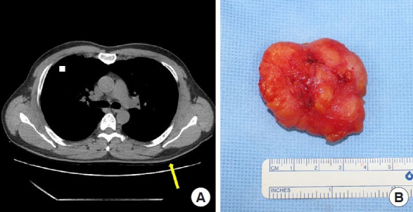
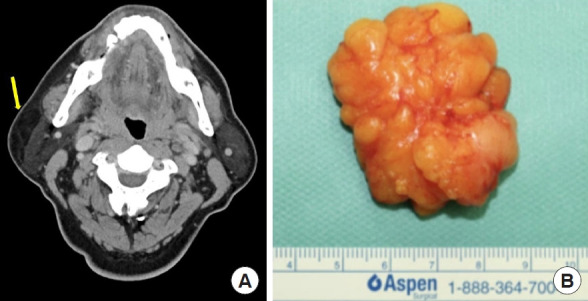
Background: Lipomas are common benign connective-tissue tumors that usually present as slow-growing, painless, subcutaneous masses. Deeper variants, such as intramuscular, intermuscular, and submuscular lipomas, are larger and rarer. Accurate preoperative depth determination is crucial for planning appropriate surgical resection.
Methods: We retrospectively reviewed 190 lipoma cases treated at a single medical center from January 2013 to August 2023. The accuracy of preoperative imaging techniques-ultrasonography (USG), computed tomography (CT), and magnetic resonance imaging (MRI)-in predicting lipoma depth was assessed.
Results: USG, CT, and MRI showed accuracies of 72.5%, 56.5%, and 79.3%, respectively, with MRI showing the highest predictive accuracy. The trunk was the most common site for lipomas (49.5%), followed by the upper (20.5%) and lower extremities (13.2%). USG was more accurate for lipomas in the lower extremities and neck, whereas CT was less accurate for lipomas in the trunk.
Conclusion: MRI is preferable for the preoperative depth assessment of lipomas, especially those located in the trunk. Accurate imaging is essential for guiding surgical planning and avoiding complications. Further studies with larger sample sizes are required to validate our findings.

 求助内容:
求助内容: 应助结果提醒方式:


