年轻雄性大鼠磨牙发育过程中根尖周病变的组织病理学评价

IF 2.2 4区 医学 Q2 DENTISTRY, ORAL SURGERY & MEDICINE
Mariana Bena Gelio , Paula Aparecida Nakazone-Guimarães , Tiago Silva da Fonseca , Evelin Carine Alves Silva , Estela Sasso-Cerri , Paulo Sérgio Cerri
{"title":"年轻雄性大鼠磨牙发育过程中根尖周病变的组织病理学评价","authors":"Mariana Bena Gelio ,&nbsp;Paula Aparecida Nakazone-Guimarães ,&nbsp;Tiago Silva da Fonseca ,&nbsp;Evelin Carine Alves Silva ,&nbsp;Estela Sasso-Cerri ,&nbsp;Paulo Sérgio Cerri","doi":"10.1016/j.archoralbio.2025.106222","DOIUrl":null,"url":null,"abstract":"<div><h3>Objectives</h3><div>To evaluate the histopathological features of induced periapical lesions (PL) exposed to the oral environment at different time points in developing molars of rats.</div></div><div><h3>Methods</h3><div>Twenty-four 30-day-old Holtzman rats had the pulp chamber of the mandibular left first molar opened and root canals were exposed to the oral environment. According to the exposure time, the animals were distributed into three groups (n = 8 rats/group): PL induced for 3 days (PLG-3d), PL induced for 1week (PLG-1w) and PL induced for 9 weeks (PLG-9w). The healthy right first molars were used as control group (CG). In paraffin-embedded jaw sections, the number of inflammatory cells (ICs), IL-6-immunolabelled cells, osteoclasts and PLs area were measured. Data were subjected to two-way ANOVA analysis of variance and Tukey's post-test (p &lt; 0.05).</div></div><div><h3>Results</h3><div>The highest values of ICs and IL-6-immunolabelled cells were found in PLG-1w while no significant difference was observed between PLG-3d and PLG-9w specimens. PLG-1w showed a significantly greater number of osteoclasts than PLG-9w, but no significant difference was found in the PL area between these groups.</div></div><div><h3>Conclusions</h3><div>Although the peak of the inflammatory infiltrate, bone resorption and PL size were reached after 1-week, periapical lesions were established after 3 days in developing molars of rats.</div></div>","PeriodicalId":8288,"journal":{"name":"Archives of oral biology","volume":"173 ","pages":"Article 106222"},"PeriodicalIF":2.2000,"publicationDate":"2025-03-05","publicationTypes":"Journal Article","fieldsOfStudy":null,"isOpenAccess":false,"openAccessPdf":"","citationCount":"0","resultStr":"{\"title\":\"Histopathological evaluation of periapical lesions in developing molars of young male rats\",\"authors\":\"Mariana Bena Gelio ,&nbsp;Paula Aparecida Nakazone-Guimarães ,&nbsp;Tiago Silva da Fonseca ,&nbsp;Evelin Carine Alves Silva ,&nbsp;Estela Sasso-Cerri ,&nbsp;Paulo Sérgio Cerri\",\"doi\":\"10.1016/j.archoralbio.2025.106222\",\"DOIUrl\":null,\"url\":null,\"abstract\":\"<div><h3>Objectives</h3><div>To evaluate the histopathological features of induced periapical lesions (PL) exposed to the oral environment at different time points in developing molars of rats.</div></div><div><h3>Methods</h3><div>Twenty-four 30-day-old Holtzman rats had the pulp chamber of the mandibular left first molar opened and root canals were exposed to the oral environment. According to the exposure time, the animals were distributed into three groups (n = 8 rats/group): PL induced for 3 days (PLG-3d), PL induced for 1week (PLG-1w) and PL induced for 9 weeks (PLG-9w). The healthy right first molars were used as control group (CG). In paraffin-embedded jaw sections, the number of inflammatory cells (ICs), IL-6-immunolabelled cells, osteoclasts and PLs area were measured. Data were subjected to two-way ANOVA analysis of variance and Tukey's post-test (p &lt; 0.05).</div></div><div><h3>Results</h3><div>The highest values of ICs and IL-6-immunolabelled cells were found in PLG-1w while no significant difference was observed between PLG-3d and PLG-9w specimens. PLG-1w showed a significantly greater number of osteoclasts than PLG-9w, but no significant difference was found in the PL area between these groups.</div></div><div><h3>Conclusions</h3><div>Although the peak of the inflammatory infiltrate, bone resorption and PL size were reached after 1-week, periapical lesions were established after 3 days in developing molars of rats.</div></div>\",\"PeriodicalId\":8288,\"journal\":{\"name\":\"Archives of oral biology\",\"volume\":\"173 \",\"pages\":\"Article 106222\"},\"PeriodicalIF\":2.2000,\"publicationDate\":\"2025-03-05\",\"publicationTypes\":\"Journal Article\",\"fieldsOfStudy\":null,\"isOpenAccess\":false,\"openAccessPdf\":\"\",\"citationCount\":\"0\",\"resultStr\":null,\"platform\":\"Semanticscholar\",\"paperid\":null,\"PeriodicalName\":\"Archives of oral biology\",\"FirstCategoryId\":\"3\",\"ListUrlMain\":\"https://www.sciencedirect.com/science/article/pii/S0003996925000500\",\"RegionNum\":4,\"RegionCategory\":\"医学\",\"ArticlePicture\":[],\"TitleCN\":null,\"AbstractTextCN\":null,\"PMCID\":null,\"EPubDate\":\"\",\"PubModel\":\"\",\"JCR\":\"Q2\",\"JCRName\":\"DENTISTRY, ORAL SURGERY & MEDICINE\",\"Score\":null,\"Total\":0}","platform":"Semanticscholar","paperid":null,"PeriodicalName":"Archives of oral biology","FirstCategoryId":"3","ListUrlMain":"https://www.sciencedirect.com/science/article/pii/S0003996925000500","RegionNum":4,"RegionCategory":"医学","ArticlePicture":[],"TitleCN":null,"AbstractTextCN":null,"PMCID":null,"EPubDate":"","PubModel":"","JCR":"Q2","JCRName":"DENTISTRY, ORAL SURGERY & MEDICINE","Score":null,"Total":0}
引用次数: 0

摘要

目的探讨大鼠磨牙发育过程中不同时间点暴露于口腔环境的诱导性根尖周病变的组织病理学特征。方法24只30日龄Holtzman大鼠,将下颌左第一磨牙牙髓室打开,根管暴露于口腔环境。根据暴露时间分为3组(n = 8只/组):PL诱导3天(PLG-3d)、PL诱导1周(PLG-1w)和PL诱导9周(PLG-9w)。以健康右侧第一磨牙为对照组(CG)。在石蜡包埋的颌骨切片中,测量炎症细胞(ic)、il -6免疫标记细胞、破骨细胞数量和PLs面积。数据采用双因素方差分析和Tukey后验(p <;0.05)。结果il -6免疫标记细胞在PLG-1w中含量最高,而在PLG-3d和PLG-9w中差异无统计学意义。PLG-1w组的破骨细胞数量明显高于PLG-9w组,但两组间PL区无明显差异。结论大鼠生长磨牙的炎症浸润、骨吸收和PL大小在1周后达到峰值,但在3天后形成根尖周病变。
本文章由计算机程序翻译,如有差异,请以英文原文为准。
Histopathological evaluation of periapical lesions in developing molars of young male rats

Objectives

To evaluate the histopathological features of induced periapical lesions (PL) exposed to the oral environment at different time points in developing molars of rats.

Methods

Twenty-four 30-day-old Holtzman rats had the pulp chamber of the mandibular left first molar opened and root canals were exposed to the oral environment. According to the exposure time, the animals were distributed into three groups (n = 8 rats/group): PL induced for 3 days (PLG-3d), PL induced for 1week (PLG-1w) and PL induced for 9 weeks (PLG-9w). The healthy right first molars were used as control group (CG). In paraffin-embedded jaw sections, the number of inflammatory cells (ICs), IL-6-immunolabelled cells, osteoclasts and PLs area were measured. Data were subjected to two-way ANOVA analysis of variance and Tukey's post-test (p < 0.05).

Results

The highest values of ICs and IL-6-immunolabelled cells were found in PLG-1w while no significant difference was observed between PLG-3d and PLG-9w specimens. PLG-1w showed a significantly greater number of osteoclasts than PLG-9w, but no significant difference was found in the PL area between these groups.

Conclusions

Although the peak of the inflammatory infiltrate, bone resorption and PL size were reached after 1-week, periapical lesions were established after 3 days in developing molars of rats.
求助全文
通过发布文献求助,成功后即可免费获取论文全文。 去求助
来源期刊
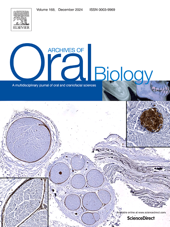
Archives of oral biology
Archives of oral biology 医学-牙科与口腔外科
CiteScore
5.10
自引率
3.30%
发文量
177
审稿时长
26 days
期刊介绍: Archives of Oral Biology is an international journal which aims to publish papers of the highest scientific quality in the oral and craniofacial sciences. The journal is particularly interested in research which advances knowledge in the mechanisms of craniofacial development and disease, including: Cell and molecular biology Molecular genetics Immunology Pathogenesis Cellular microbiology Embryology Syndromology Forensic dentistry
×
引用
GB/T 7714-2015
复制
MLA
复制
APA
复制
导出至
BibTeX EndNote RefMan NoteFirst NoteExpress
×
提示
您的信息不完整,为了账户安全,请先补充。
现在去补充
×
提示
您因"违规操作"
具体请查看互助需知
我知道了
×
提示
确定
请完成安全验证×
copy
已复制链接
快去分享给好友吧!
我知道了
右上角分享
点击右上角分享
0
联系我们:info@booksci.cn Book学术提供免费学术资源搜索服务,方便国内外学者检索中英文文献。致力于提供最便捷和优质的服务体验。 Copyright © 2023 布克学术 All rights reserved.
京ICP备2023020795号-1
ghs 京公网安备 11010802042870号
Book学术文献互助
Book学术文献互助群
群 号:481959085
Book学术官方微信