Koichiro M. Hirosawa, Yusuke Sato, Rinshi S. Kasai, Eriko Yamaguchi, Naoko Komura, Hiromune Ando, Ayuko Hoshino, Yasunari Yokota, Kenichi G. N. Suzuki
{"title":"受体细胞摄取小的细胞外囊泡是由旁分泌粘连信号促进的","authors":"Koichiro M. Hirosawa, Yusuke Sato, Rinshi S. Kasai, Eriko Yamaguchi, Naoko Komura, Hiromune Ando, Ayuko Hoshino, Yasunari Yokota, Kenichi G. N. Suzuki","doi":"10.1038/s41467-025-57617-9","DOIUrl":null,"url":null,"abstract":"<p>Small extracellular vesicles (sEVs) play crucial roles in intercellular communication. However, the internalization of individual sEVs by recipient cells has not been directly observed. Here, we examined these mechanisms using state-of-the-art imaging techniques. Single-molecule imaging shows that tumor-derived sEVs can be classified into several subtypes. Simultaneous single-sEV particle tracking and observation of super-resolution movies of membrane invaginations in living cells reveal that all sEV subtypes are internalized via clathrin-independent endocytosis mediated by galectin-3 and lysosome-associated membrane protein-2C, while some subtypes that recruited raft markers are internalized through caveolae. Integrin β1 and talin-1 accumulate in recipient cell plasma membranes beneath all sEV subtypes. Paracrine, but not autocrine, sEV binding triggers Ca<sup>2+</sup> mobilization induced by the activation of Src family kinases and phospholipase Cγ. Subsequent Ca<sup>2+</sup>-induced activation of calcineurin–dynamin promotes sEV internalization, leading to the recycling pathway. Thus, we clarified the detailed mechanisms of sEV internalization driven by paracrine adhesion signaling.</p>","PeriodicalId":19066,"journal":{"name":"Nature Communications","volume":"56 1","pages":""},"PeriodicalIF":15.7000,"publicationDate":"2025-03-12","publicationTypes":"Journal Article","fieldsOfStudy":null,"isOpenAccess":false,"openAccessPdf":"","citationCount":"0","resultStr":"{\"title\":\"Uptake of small extracellular vesicles by recipient cells is facilitated by paracrine adhesion signaling\",\"authors\":\"Koichiro M. Hirosawa, Yusuke Sato, Rinshi S. Kasai, Eriko Yamaguchi, Naoko Komura, Hiromune Ando, Ayuko Hoshino, Yasunari Yokota, Kenichi G. N. Suzuki\",\"doi\":\"10.1038/s41467-025-57617-9\",\"DOIUrl\":null,\"url\":null,\"abstract\":\"<p>Small extracellular vesicles (sEVs) play crucial roles in intercellular communication. However, the internalization of individual sEVs by recipient cells has not been directly observed. Here, we examined these mechanisms using state-of-the-art imaging techniques. Single-molecule imaging shows that tumor-derived sEVs can be classified into several subtypes. Simultaneous single-sEV particle tracking and observation of super-resolution movies of membrane invaginations in living cells reveal that all sEV subtypes are internalized via clathrin-independent endocytosis mediated by galectin-3 and lysosome-associated membrane protein-2C, while some subtypes that recruited raft markers are internalized through caveolae. Integrin β1 and talin-1 accumulate in recipient cell plasma membranes beneath all sEV subtypes. Paracrine, but not autocrine, sEV binding triggers Ca<sup>2+</sup> mobilization induced by the activation of Src family kinases and phospholipase Cγ. Subsequent Ca<sup>2+</sup>-induced activation of calcineurin–dynamin promotes sEV internalization, leading to the recycling pathway. Thus, we clarified the detailed mechanisms of sEV internalization driven by paracrine adhesion signaling.</p>\",\"PeriodicalId\":19066,\"journal\":{\"name\":\"Nature Communications\",\"volume\":\"56 1\",\"pages\":\"\"},\"PeriodicalIF\":15.7000,\"publicationDate\":\"2025-03-12\",\"publicationTypes\":\"Journal Article\",\"fieldsOfStudy\":null,\"isOpenAccess\":false,\"openAccessPdf\":\"\",\"citationCount\":\"0\",\"resultStr\":null,\"platform\":\"Semanticscholar\",\"paperid\":null,\"PeriodicalName\":\"Nature Communications\",\"FirstCategoryId\":\"103\",\"ListUrlMain\":\"https://doi.org/10.1038/s41467-025-57617-9\",\"RegionNum\":1,\"RegionCategory\":\"综合性期刊\",\"ArticlePicture\":[],\"TitleCN\":null,\"AbstractTextCN\":null,\"PMCID\":null,\"EPubDate\":\"\",\"PubModel\":\"\",\"JCR\":\"Q1\",\"JCRName\":\"MULTIDISCIPLINARY SCIENCES\",\"Score\":null,\"Total\":0}","platform":"Semanticscholar","paperid":null,"PeriodicalName":"Nature Communications","FirstCategoryId":"103","ListUrlMain":"https://doi.org/10.1038/s41467-025-57617-9","RegionNum":1,"RegionCategory":"综合性期刊","ArticlePicture":[],"TitleCN":null,"AbstractTextCN":null,"PMCID":null,"EPubDate":"","PubModel":"","JCR":"Q1","JCRName":"MULTIDISCIPLINARY SCIENCES","Score":null,"Total":0}
Uptake of small extracellular vesicles by recipient cells is facilitated by paracrine adhesion signaling
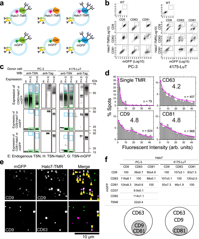
Small extracellular vesicles (sEVs) play crucial roles in intercellular communication. However, the internalization of individual sEVs by recipient cells has not been directly observed. Here, we examined these mechanisms using state-of-the-art imaging techniques. Single-molecule imaging shows that tumor-derived sEVs can be classified into several subtypes. Simultaneous single-sEV particle tracking and observation of super-resolution movies of membrane invaginations in living cells reveal that all sEV subtypes are internalized via clathrin-independent endocytosis mediated by galectin-3 and lysosome-associated membrane protein-2C, while some subtypes that recruited raft markers are internalized through caveolae. Integrin β1 and talin-1 accumulate in recipient cell plasma membranes beneath all sEV subtypes. Paracrine, but not autocrine, sEV binding triggers Ca2+ mobilization induced by the activation of Src family kinases and phospholipase Cγ. Subsequent Ca2+-induced activation of calcineurin–dynamin promotes sEV internalization, leading to the recycling pathway. Thus, we clarified the detailed mechanisms of sEV internalization driven by paracrine adhesion signaling.
期刊介绍:
Nature Communications, an open-access journal, publishes high-quality research spanning all areas of the natural sciences. Papers featured in the journal showcase significant advances relevant to specialists in each respective field. With a 2-year impact factor of 16.6 (2022) and a median time of 8 days from submission to the first editorial decision, Nature Communications is committed to rapid dissemination of research findings. As a multidisciplinary journal, it welcomes contributions from biological, health, physical, chemical, Earth, social, mathematical, applied, and engineering sciences, aiming to highlight important breakthroughs within each domain.
 求助内容:
求助内容: 应助结果提醒方式:


