Dan Mocanu, Katarzyna Bokwa-Dąbrowska, Katarina Nilsson Helander, Pawel Szaro
{"title":"超声与磁共振成像诊断踝关节后外侧疼痛的比较分析。","authors":"Dan Mocanu, Katarzyna Bokwa-Dąbrowska, Katarina Nilsson Helander, Pawel Szaro","doi":"10.15557/JoU.2025.0002","DOIUrl":null,"url":null,"abstract":"<p><strong>Aim: </strong>The purpose of this study was to evaluate the diagnostic value of ultrasound compared to magnetic resonance imaging (MRI) as a reference in detecting peroneus brevis split ruptures.</p><p><strong>Material and methods: </strong>We re-reviewed 112 ultrasound examinations performed between 2020 and 2021 by three musculoskeletal radiologists with 8-10 years of experience. Patients were referred due to pain lasting at least 8 months in the posterolateral ankle. Ultrasound was performed using a LOGIQ E9 General Electric device with a 6-15 MHz or 18 MHz probe. Sixty-three patients who underwent MRI within 8 months and were included in the study. Ultrasound and MRI findings were categorized as: a) no peroneus split, b) presence of peroneus split, or c) unspecific findings. MRI served as the reference standard. Sensitivity, specificity, positive predictive value, negative predictive value, and accuracy were calculated.</p><p><strong>Results: </strong>Seven cases (11.1%) were false positives (diagnosed on ultrasound but not MRI) and 9 (14.3%) were false negatives (missed by ultrasound but detected on MRI). Six cases (9.5%) were true positives (identified on both ultrasound and MRI), and 41 patients (65.1%) were true negatives (negative on both modalities). Ultrasound showed a sensitivity of 40.0% and specificity of 85.4%. The positive predictive value (PPV) was 46.2%, while the negative predictive value (NPV) was 82.0%.</p><p><strong>Conclusions: </strong>Ultrasound demonstrated limited sensitivity but high specificity in detecting peroneus brevis split ruptures.</p>","PeriodicalId":45612,"journal":{"name":"Journal of Ultrasonography","volume":"25 100","pages":"20250002"},"PeriodicalIF":1.5000,"publicationDate":"2025-01-23","publicationTypes":"Journal Article","fieldsOfStudy":null,"isOpenAccess":false,"openAccessPdf":"https://www.ncbi.nlm.nih.gov/pmc/articles/PMC11893017/pdf/","citationCount":"0","resultStr":"{\"title\":\"Comparative analysis of ultrasound and magnetic resonance imaging in diagnosing pain in the posterolateral region of the ankle.\",\"authors\":\"Dan Mocanu, Katarzyna Bokwa-Dąbrowska, Katarina Nilsson Helander, Pawel Szaro\",\"doi\":\"10.15557/JoU.2025.0002\",\"DOIUrl\":null,\"url\":null,\"abstract\":\"<p><strong>Aim: </strong>The purpose of this study was to evaluate the diagnostic value of ultrasound compared to magnetic resonance imaging (MRI) as a reference in detecting peroneus brevis split ruptures.</p><p><strong>Material and methods: </strong>We re-reviewed 112 ultrasound examinations performed between 2020 and 2021 by three musculoskeletal radiologists with 8-10 years of experience. Patients were referred due to pain lasting at least 8 months in the posterolateral ankle. Ultrasound was performed using a LOGIQ E9 General Electric device with a 6-15 MHz or 18 MHz probe. Sixty-three patients who underwent MRI within 8 months and were included in the study. Ultrasound and MRI findings were categorized as: a) no peroneus split, b) presence of peroneus split, or c) unspecific findings. MRI served as the reference standard. Sensitivity, specificity, positive predictive value, negative predictive value, and accuracy were calculated.</p><p><strong>Results: </strong>Seven cases (11.1%) were false positives (diagnosed on ultrasound but not MRI) and 9 (14.3%) were false negatives (missed by ultrasound but detected on MRI). Six cases (9.5%) were true positives (identified on both ultrasound and MRI), and 41 patients (65.1%) were true negatives (negative on both modalities). Ultrasound showed a sensitivity of 40.0% and specificity of 85.4%. The positive predictive value (PPV) was 46.2%, while the negative predictive value (NPV) was 82.0%.</p><p><strong>Conclusions: </strong>Ultrasound demonstrated limited sensitivity but high specificity in detecting peroneus brevis split ruptures.</p>\",\"PeriodicalId\":45612,\"journal\":{\"name\":\"Journal of Ultrasonography\",\"volume\":\"25 100\",\"pages\":\"20250002\"},\"PeriodicalIF\":1.5000,\"publicationDate\":\"2025-01-23\",\"publicationTypes\":\"Journal Article\",\"fieldsOfStudy\":null,\"isOpenAccess\":false,\"openAccessPdf\":\"https://www.ncbi.nlm.nih.gov/pmc/articles/PMC11893017/pdf/\",\"citationCount\":\"0\",\"resultStr\":null,\"platform\":\"Semanticscholar\",\"paperid\":null,\"PeriodicalName\":\"Journal of Ultrasonography\",\"FirstCategoryId\":\"1085\",\"ListUrlMain\":\"https://doi.org/10.15557/JoU.2025.0002\",\"RegionNum\":0,\"RegionCategory\":null,\"ArticlePicture\":[],\"TitleCN\":null,\"AbstractTextCN\":null,\"PMCID\":null,\"EPubDate\":\"2025/1/1 0:00:00\",\"PubModel\":\"eCollection\",\"JCR\":\"Q3\",\"JCRName\":\"RADIOLOGY, NUCLEAR MEDICINE & MEDICAL IMAGING\",\"Score\":null,\"Total\":0}","platform":"Semanticscholar","paperid":null,"PeriodicalName":"Journal of Ultrasonography","FirstCategoryId":"1085","ListUrlMain":"https://doi.org/10.15557/JoU.2025.0002","RegionNum":0,"RegionCategory":null,"ArticlePicture":[],"TitleCN":null,"AbstractTextCN":null,"PMCID":null,"EPubDate":"2025/1/1 0:00:00","PubModel":"eCollection","JCR":"Q3","JCRName":"RADIOLOGY, NUCLEAR MEDICINE & MEDICAL IMAGING","Score":null,"Total":0}
Comparative analysis of ultrasound and magnetic resonance imaging in diagnosing pain in the posterolateral region of the ankle.
Aim: The purpose of this study was to evaluate the diagnostic value of ultrasound compared to magnetic resonance imaging (MRI) as a reference in detecting peroneus brevis split ruptures.
Material and methods: We re-reviewed 112 ultrasound examinations performed between 2020 and 2021 by three musculoskeletal radiologists with 8-10 years of experience. Patients were referred due to pain lasting at least 8 months in the posterolateral ankle. Ultrasound was performed using a LOGIQ E9 General Electric device with a 6-15 MHz or 18 MHz probe. Sixty-three patients who underwent MRI within 8 months and were included in the study. Ultrasound and MRI findings were categorized as: a) no peroneus split, b) presence of peroneus split, or c) unspecific findings. MRI served as the reference standard. Sensitivity, specificity, positive predictive value, negative predictive value, and accuracy were calculated.
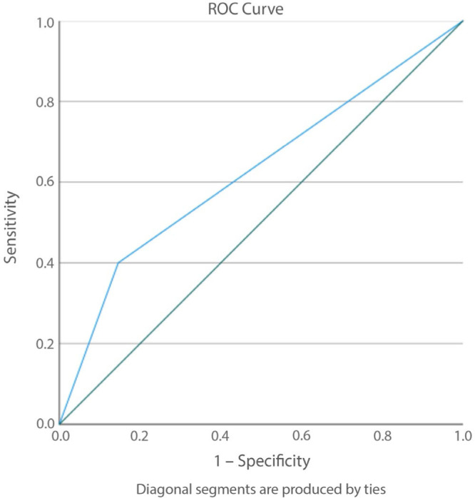
Results: Seven cases (11.1%) were false positives (diagnosed on ultrasound but not MRI) and 9 (14.3%) were false negatives (missed by ultrasound but detected on MRI). Six cases (9.5%) were true positives (identified on both ultrasound and MRI), and 41 patients (65.1%) were true negatives (negative on both modalities). Ultrasound showed a sensitivity of 40.0% and specificity of 85.4%. The positive predictive value (PPV) was 46.2%, while the negative predictive value (NPV) was 82.0%.
Conclusions: Ultrasound demonstrated limited sensitivity but high specificity in detecting peroneus brevis split ruptures.

 求助内容:
求助内容: 应助结果提醒方式:


