Ni Chen, Hanlin Liang, Siqiao Liang, Xiaona Liang, Xuemei Huang, Qingliang Yu, Zhiyi He
{"title":"血清 IgE 与抗干扰素-γ 自身抗体综合征的临床特征和疾病预后的关系。","authors":"Ni Chen, Hanlin Liang, Siqiao Liang, Xiaona Liang, Xuemei Huang, Qingliang Yu, Zhiyi He","doi":"10.1186/s12865-025-00696-6","DOIUrl":null,"url":null,"abstract":"<p><strong>Background: </strong>Anti-interferon-γ autoantibodies (AIGAs) syndrome is a recently recognized adult-onset immunodeficiency syndrome. Serum Immunoglobulin E (IgE) is increased in AIGAs syndrome, but the role of serum IgE levels in the clinical features and disease outcomes of AIGAs syndrome is not clear.</p><p><strong>Methods: </strong>We retrospectively enrolled 163 patients diagnosed AIGAs syndrome with serum IgE examined at baseline from 2021 to 2024 and compared the clinical features between Group A (serum IgE level ≤ 212 IU/mL) and Group B (serum IgE level > 212 IU/mL). Multivariable logistic regression method was used to explore the risk factors associated with disease outcomes.</p><p><strong>Results: </strong>163 patients were included in this study, of whom 97 patients were in Group A (serum IgE level ≤ 212 IU/mL) and 66 patients in Group B (serum IgE level > 212 IU/mL). Group B showed higher number of infectious episodes, elevated levels of erythrocyte sedimentation rate (ESR), CD3 + T cells, immunoglobulin G (IgG), IgA, and globulins (GLB), shorter progression-free survival (PFS), and increased exacerbation numbers. Group B exhibited a higher incidence of fatigue, dyspnea, loss of appetite, rash, moist rales, hepatomegaly, and splenomegaly. Skin, bone marrow and spleen involvements were more common in Group B. IgE demonstrated correlations with IgG, GLB, Albumin (ALB), Eosinophils (EOS), IgG4, and ESR. During the follow-up, Group B exhibiting higher number of exacerbations compared to Group A (P < 0.0001). Multivariable Cox regression analysis revealed that High AIGAs titers (hazard ratio [HR], 2.418, 95% confidence interval [CI]1.037-5.642, P = 0.041), WBC > 22.52 × 10<sup>9</sup>cells/L (HR2.199, 95%CI1.194-4.050, P = 0.012) were independent risk factors of disease exacerbation. Glucocorticoid treatment was commonly used in patients with AIGAs syndrome who had elevated IgE levels and skin involvement, demonstrating efficacy in improving condition.</p><p><strong>Conclusions: </strong>Elevated serum IgE levels are associated with more severe clinical features in AIGAs syndrome, including increased infectious episodes, elevated inflammatory markers/immune markers, and multi-organ involvement, particularly skin. IgE serves as a marker of skin involvement and may indicate a potential response to glucocorticoid treatment.</p>","PeriodicalId":9040,"journal":{"name":"BMC Immunology","volume":"26 1","pages":"17"},"PeriodicalIF":2.7000,"publicationDate":"2025-03-08","publicationTypes":"Journal Article","fieldsOfStudy":null,"isOpenAccess":false,"openAccessPdf":"https://www.ncbi.nlm.nih.gov/pmc/articles/PMC11889914/pdf/","citationCount":"0","resultStr":"{\"title\":\"Serum IgE in the clinical features and disease outcomes of anti-interferon-γ autoantibodies syndrome.\",\"authors\":\"Ni Chen, Hanlin Liang, Siqiao Liang, Xiaona Liang, Xuemei Huang, Qingliang Yu, Zhiyi He\",\"doi\":\"10.1186/s12865-025-00696-6\",\"DOIUrl\":null,\"url\":null,\"abstract\":\"<p><strong>Background: </strong>Anti-interferon-γ autoantibodies (AIGAs) syndrome is a recently recognized adult-onset immunodeficiency syndrome. Serum Immunoglobulin E (IgE) is increased in AIGAs syndrome, but the role of serum IgE levels in the clinical features and disease outcomes of AIGAs syndrome is not clear.</p><p><strong>Methods: </strong>We retrospectively enrolled 163 patients diagnosed AIGAs syndrome with serum IgE examined at baseline from 2021 to 2024 and compared the clinical features between Group A (serum IgE level ≤ 212 IU/mL) and Group B (serum IgE level > 212 IU/mL). Multivariable logistic regression method was used to explore the risk factors associated with disease outcomes.</p><p><strong>Results: </strong>163 patients were included in this study, of whom 97 patients were in Group A (serum IgE level ≤ 212 IU/mL) and 66 patients in Group B (serum IgE level > 212 IU/mL). Group B showed higher number of infectious episodes, elevated levels of erythrocyte sedimentation rate (ESR), CD3 + T cells, immunoglobulin G (IgG), IgA, and globulins (GLB), shorter progression-free survival (PFS), and increased exacerbation numbers. Group B exhibited a higher incidence of fatigue, dyspnea, loss of appetite, rash, moist rales, hepatomegaly, and splenomegaly. Skin, bone marrow and spleen involvements were more common in Group B. IgE demonstrated correlations with IgG, GLB, Albumin (ALB), Eosinophils (EOS), IgG4, and ESR. During the follow-up, Group B exhibiting higher number of exacerbations compared to Group A (P < 0.0001). Multivariable Cox regression analysis revealed that High AIGAs titers (hazard ratio [HR], 2.418, 95% confidence interval [CI]1.037-5.642, P = 0.041), WBC > 22.52 × 10<sup>9</sup>cells/L (HR2.199, 95%CI1.194-4.050, P = 0.012) were independent risk factors of disease exacerbation. Glucocorticoid treatment was commonly used in patients with AIGAs syndrome who had elevated IgE levels and skin involvement, demonstrating efficacy in improving condition.</p><p><strong>Conclusions: </strong>Elevated serum IgE levels are associated with more severe clinical features in AIGAs syndrome, including increased infectious episodes, elevated inflammatory markers/immune markers, and multi-organ involvement, particularly skin. IgE serves as a marker of skin involvement and may indicate a potential response to glucocorticoid treatment.</p>\",\"PeriodicalId\":9040,\"journal\":{\"name\":\"BMC Immunology\",\"volume\":\"26 1\",\"pages\":\"17\"},\"PeriodicalIF\":2.7000,\"publicationDate\":\"2025-03-08\",\"publicationTypes\":\"Journal Article\",\"fieldsOfStudy\":null,\"isOpenAccess\":false,\"openAccessPdf\":\"https://www.ncbi.nlm.nih.gov/pmc/articles/PMC11889914/pdf/\",\"citationCount\":\"0\",\"resultStr\":null,\"platform\":\"Semanticscholar\",\"paperid\":null,\"PeriodicalName\":\"BMC Immunology\",\"FirstCategoryId\":\"3\",\"ListUrlMain\":\"https://doi.org/10.1186/s12865-025-00696-6\",\"RegionNum\":4,\"RegionCategory\":\"医学\",\"ArticlePicture\":[],\"TitleCN\":null,\"AbstractTextCN\":null,\"PMCID\":null,\"EPubDate\":\"\",\"PubModel\":\"\",\"JCR\":\"Q3\",\"JCRName\":\"IMMUNOLOGY\",\"Score\":null,\"Total\":0}","platform":"Semanticscholar","paperid":null,"PeriodicalName":"BMC Immunology","FirstCategoryId":"3","ListUrlMain":"https://doi.org/10.1186/s12865-025-00696-6","RegionNum":4,"RegionCategory":"医学","ArticlePicture":[],"TitleCN":null,"AbstractTextCN":null,"PMCID":null,"EPubDate":"","PubModel":"","JCR":"Q3","JCRName":"IMMUNOLOGY","Score":null,"Total":0}
Serum IgE in the clinical features and disease outcomes of anti-interferon-γ autoantibodies syndrome.
Background: Anti-interferon-γ autoantibodies (AIGAs) syndrome is a recently recognized adult-onset immunodeficiency syndrome. Serum Immunoglobulin E (IgE) is increased in AIGAs syndrome, but the role of serum IgE levels in the clinical features and disease outcomes of AIGAs syndrome is not clear.
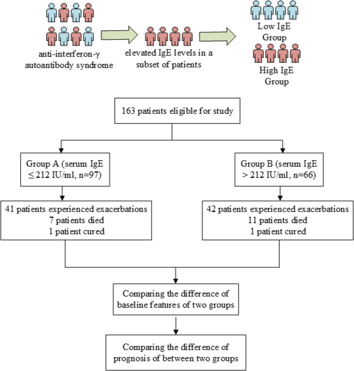
Methods: We retrospectively enrolled 163 patients diagnosed AIGAs syndrome with serum IgE examined at baseline from 2021 to 2024 and compared the clinical features between Group A (serum IgE level ≤ 212 IU/mL) and Group B (serum IgE level > 212 IU/mL). Multivariable logistic regression method was used to explore the risk factors associated with disease outcomes.
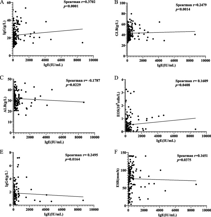
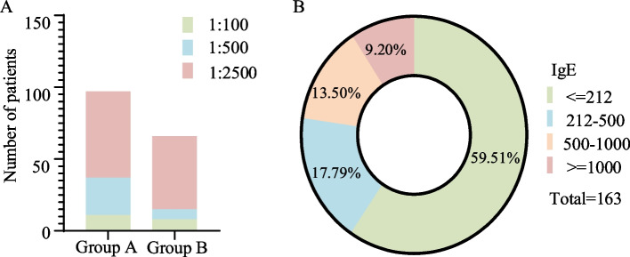
Results: 163 patients were included in this study, of whom 97 patients were in Group A (serum IgE level ≤ 212 IU/mL) and 66 patients in Group B (serum IgE level > 212 IU/mL). Group B showed higher number of infectious episodes, elevated levels of erythrocyte sedimentation rate (ESR), CD3 + T cells, immunoglobulin G (IgG), IgA, and globulins (GLB), shorter progression-free survival (PFS), and increased exacerbation numbers. Group B exhibited a higher incidence of fatigue, dyspnea, loss of appetite, rash, moist rales, hepatomegaly, and splenomegaly. Skin, bone marrow and spleen involvements were more common in Group B. IgE demonstrated correlations with IgG, GLB, Albumin (ALB), Eosinophils (EOS), IgG4, and ESR. During the follow-up, Group B exhibiting higher number of exacerbations compared to Group A (P < 0.0001). Multivariable Cox regression analysis revealed that High AIGAs titers (hazard ratio [HR], 2.418, 95% confidence interval [CI]1.037-5.642, P = 0.041), WBC > 22.52 × 10⁹cells/L (HR2.199, 95%CI1.194-4.050, P = 0.012) were independent risk factors of disease exacerbation. Glucocorticoid treatment was commonly used in patients with AIGAs syndrome who had elevated IgE levels and skin involvement, demonstrating efficacy in improving condition.
Conclusions: Elevated serum IgE levels are associated with more severe clinical features in AIGAs syndrome, including increased infectious episodes, elevated inflammatory markers/immune markers, and multi-organ involvement, particularly skin. IgE serves as a marker of skin involvement and may indicate a potential response to glucocorticoid treatment.
期刊介绍:
BMC Immunology is an open access journal publishing original peer-reviewed research articles in molecular, cellular, tissue-level, organismal, functional, and developmental aspects of the immune system as well as clinical studies and animal models of human diseases.
 求助内容:
求助内容: 应助结果提醒方式:


