Yan Zhu, Li Wang, Aichao Ruan, Zhiyu Peng, Zhenzhong Zhang
{"title":"基于放射组学和机器学习的胸腺肿块术前多分类。","authors":"Yan Zhu, Li Wang, Aichao Ruan, Zhiyu Peng, Zhenzhong Zhang","doi":"10.1186/s40644-025-00839-3","DOIUrl":null,"url":null,"abstract":"<p><strong>Background: </strong>Apart from rare cases such as lymphomas, germ cell tumors, neuroendocrine neoplasms, and thymic hyperplasia, thymic mass lesions (TMLs) are typically categorized into cysts, and thymomas. However, the classification results cannot be determined in advance and can only be confirmed through postoperative pathology. Therefore, the objective of this study is to rely on clinical parameters and radiomic features extracted from chest computed tomography (CT) scans to facilitate the preoperative classification of TMLs. The model development specifically focused on thymic cysts and thymomas, as these are the most commonly encountered anterior mediastinal tumors in clinical practice.</p><p><strong>Materials and methods: </strong>This retrospective study included 400 participants from 3 hospitals between September 2017 and September 2024 due to TMLs. The participants were classified into 7 groups based on the ultimately confirmed etiology: thymic cysts and thymomas, including types A, AB, B1, B2, B3, and C. All participants underwent contrast-enhanced chest CT scans, with senior radiologists delineating regions of interest to extract radiomic features. Additionally, the participants' ages were also collected as clinical parameters for analysis. The participants were randomly allocated into a training set and a validation set at a 7:3 ratio. A classifier models were developed using the data from the training set, and their performances were evaluated on the validation set.</p><p><strong>Results: </strong>The model exhibited good classification performance with accuracies of 0.8547.</p><p><strong>Conclusion: </strong>The model can assist in early diagnosis and the development of personalized treatment strategies for patients with TMLs.</p>","PeriodicalId":9548,"journal":{"name":"Cancer Imaging","volume":"25 1","pages":"25"},"PeriodicalIF":3.5000,"publicationDate":"2025-03-06","publicationTypes":"Journal Article","fieldsOfStudy":null,"isOpenAccess":false,"openAccessPdf":"https://www.ncbi.nlm.nih.gov/pmc/articles/PMC11884038/pdf/","citationCount":"0","resultStr":"{\"title\":\"Preoperative multiclass classification of thymic mass lesions based on radiomics and machine learning.\",\"authors\":\"Yan Zhu, Li Wang, Aichao Ruan, Zhiyu Peng, Zhenzhong Zhang\",\"doi\":\"10.1186/s40644-025-00839-3\",\"DOIUrl\":null,\"url\":null,\"abstract\":\"<p><strong>Background: </strong>Apart from rare cases such as lymphomas, germ cell tumors, neuroendocrine neoplasms, and thymic hyperplasia, thymic mass lesions (TMLs) are typically categorized into cysts, and thymomas. However, the classification results cannot be determined in advance and can only be confirmed through postoperative pathology. Therefore, the objective of this study is to rely on clinical parameters and radiomic features extracted from chest computed tomography (CT) scans to facilitate the preoperative classification of TMLs. The model development specifically focused on thymic cysts and thymomas, as these are the most commonly encountered anterior mediastinal tumors in clinical practice.</p><p><strong>Materials and methods: </strong>This retrospective study included 400 participants from 3 hospitals between September 2017 and September 2024 due to TMLs. The participants were classified into 7 groups based on the ultimately confirmed etiology: thymic cysts and thymomas, including types A, AB, B1, B2, B3, and C. All participants underwent contrast-enhanced chest CT scans, with senior radiologists delineating regions of interest to extract radiomic features. Additionally, the participants' ages were also collected as clinical parameters for analysis. The participants were randomly allocated into a training set and a validation set at a 7:3 ratio. A classifier models were developed using the data from the training set, and their performances were evaluated on the validation set.</p><p><strong>Results: </strong>The model exhibited good classification performance with accuracies of 0.8547.</p><p><strong>Conclusion: </strong>The model can assist in early diagnosis and the development of personalized treatment strategies for patients with TMLs.</p>\",\"PeriodicalId\":9548,\"journal\":{\"name\":\"Cancer Imaging\",\"volume\":\"25 1\",\"pages\":\"25\"},\"PeriodicalIF\":3.5000,\"publicationDate\":\"2025-03-06\",\"publicationTypes\":\"Journal Article\",\"fieldsOfStudy\":null,\"isOpenAccess\":false,\"openAccessPdf\":\"https://www.ncbi.nlm.nih.gov/pmc/articles/PMC11884038/pdf/\",\"citationCount\":\"0\",\"resultStr\":null,\"platform\":\"Semanticscholar\",\"paperid\":null,\"PeriodicalName\":\"Cancer Imaging\",\"FirstCategoryId\":\"3\",\"ListUrlMain\":\"https://doi.org/10.1186/s40644-025-00839-3\",\"RegionNum\":2,\"RegionCategory\":\"医学\",\"ArticlePicture\":[],\"TitleCN\":null,\"AbstractTextCN\":null,\"PMCID\":null,\"EPubDate\":\"\",\"PubModel\":\"\",\"JCR\":\"Q2\",\"JCRName\":\"ONCOLOGY\",\"Score\":null,\"Total\":0}","platform":"Semanticscholar","paperid":null,"PeriodicalName":"Cancer Imaging","FirstCategoryId":"3","ListUrlMain":"https://doi.org/10.1186/s40644-025-00839-3","RegionNum":2,"RegionCategory":"医学","ArticlePicture":[],"TitleCN":null,"AbstractTextCN":null,"PMCID":null,"EPubDate":"","PubModel":"","JCR":"Q2","JCRName":"ONCOLOGY","Score":null,"Total":0}
Preoperative multiclass classification of thymic mass lesions based on radiomics and machine learning.
Background: Apart from rare cases such as lymphomas, germ cell tumors, neuroendocrine neoplasms, and thymic hyperplasia, thymic mass lesions (TMLs) are typically categorized into cysts, and thymomas. However, the classification results cannot be determined in advance and can only be confirmed through postoperative pathology. Therefore, the objective of this study is to rely on clinical parameters and radiomic features extracted from chest computed tomography (CT) scans to facilitate the preoperative classification of TMLs. The model development specifically focused on thymic cysts and thymomas, as these are the most commonly encountered anterior mediastinal tumors in clinical practice.
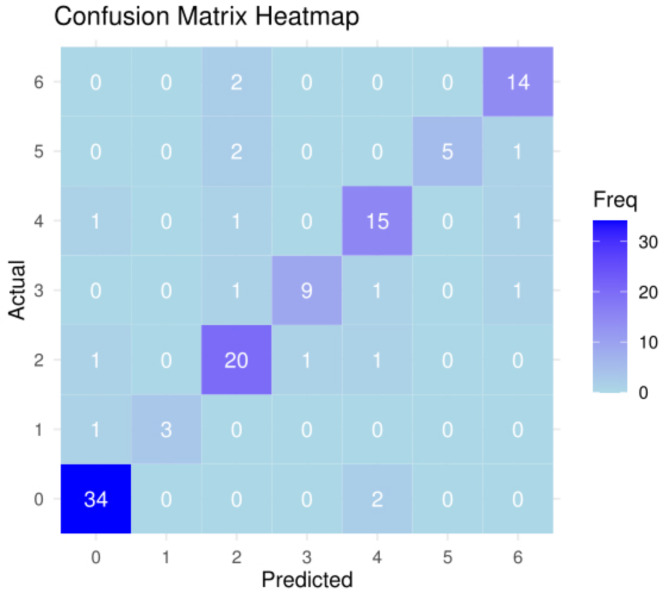
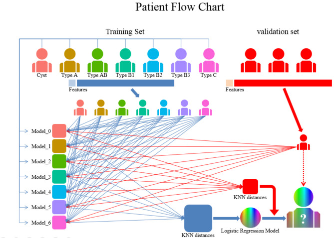
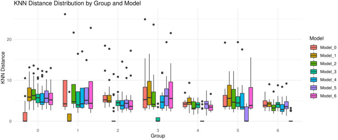
Materials and methods: This retrospective study included 400 participants from 3 hospitals between September 2017 and September 2024 due to TMLs. The participants were classified into 7 groups based on the ultimately confirmed etiology: thymic cysts and thymomas, including types A, AB, B1, B2, B3, and C. All participants underwent contrast-enhanced chest CT scans, with senior radiologists delineating regions of interest to extract radiomic features. Additionally, the participants' ages were also collected as clinical parameters for analysis. The participants were randomly allocated into a training set and a validation set at a 7:3 ratio. A classifier models were developed using the data from the training set, and their performances were evaluated on the validation set.
Results: The model exhibited good classification performance with accuracies of 0.8547.
Conclusion: The model can assist in early diagnosis and the development of personalized treatment strategies for patients with TMLs.
Cancer ImagingONCOLOGY-RADIOLOGY, NUCLEAR MEDICINE & MEDICAL IMAGING
CiteScore
7.00
自引率
0.00%
发文量
66
审稿时长
>12 weeks
期刊介绍:
Cancer Imaging is an open access, peer-reviewed journal publishing original articles, reviews and editorials written by expert international radiologists working in oncology.
The journal encompasses CT, MR, PET, ultrasound, radionuclide and multimodal imaging in all kinds of malignant tumours, plus new developments, techniques and innovations. Topics of interest include:
Breast Imaging
Chest
Complications of treatment
Ear, Nose & Throat
Gastrointestinal
Hepatobiliary & Pancreatic
Imaging biomarkers
Interventional
Lymphoma
Measurement of tumour response
Molecular functional imaging
Musculoskeletal
Neuro oncology
Nuclear Medicine
Paediatric.

 求助内容:
求助内容: 应助结果提醒方式:


