{"title":"腹内侧前额叶皮层突出的团聚核神经元在厌恶记忆消退过程中的不同神经反应。","authors":"Yuki Mochizuki, Asuka Joji-Nishino, Kazuo Emoto, Akira Uematsu","doi":"10.1186/s13041-025-01185-y","DOIUrl":null,"url":null,"abstract":"<p><p>Animals adaptively regulate aversive memories in safe environments through extinction, a process central to exposure therapy for anxiety disorders. The limbic thalamus controls cognitive function in concert with interconnected cortical and limbic structures. Though medial prefrontal (mPFC) afferents to the limbic thalamus regulate aversive memory, the functional role of limbic thalamus efferents to mPFC is unclear. Here, we investigated the roles of thalamic nuclei, the reuniens (RE) and mediodorsal (MD) thalamus, projecting to the medial prefrontal cortex (mPFC) in aversive memory conditioning and extinction in male mice. Using retrograde tracing, we demonstrated that ventromedial PFC (vmPFC)- and dorsomedial PFC (dmPFC)-projecting neurons are topologically segregated within the RE and MD. Fiber photometry revealed that both RE→vmPFC and MD→vmPFC neurons respond to aversive stimuli. Notably, RE→vmPFC neurons develop shock-associated cue (CS+) response during aversive conditioning. During extinction, RE→vmPFC neurons exhibited a biphasic response to CS+, while MD→vmPFC neurons showed no cue-evoked activity. Neither optogenetic activation nor inactivation of these populations altered freezing behavior during extinction compared to controls. Collectively, these findings indicate that RE→vmPFC neurons encode aversive cue information during extinction but are dispensable for behavioral modulation. This study highlights the distinct contributions of limbic thalamus-PFC circuits to aversive memory processing.</p>","PeriodicalId":18851,"journal":{"name":"Molecular Brain","volume":"18 1","pages":"18"},"PeriodicalIF":2.9000,"publicationDate":"2025-03-05","publicationTypes":"Journal Article","fieldsOfStudy":null,"isOpenAccess":false,"openAccessPdf":"https://www.ncbi.nlm.nih.gov/pmc/articles/PMC11881366/pdf/","citationCount":"0","resultStr":"{\"title\":\"Distinct neural responses of ventromedial prefrontal cortex-projecting nucleus reuniens neurons during aversive memory extinction.\",\"authors\":\"Yuki Mochizuki, Asuka Joji-Nishino, Kazuo Emoto, Akira Uematsu\",\"doi\":\"10.1186/s13041-025-01185-y\",\"DOIUrl\":null,\"url\":null,\"abstract\":\"<p><p>Animals adaptively regulate aversive memories in safe environments through extinction, a process central to exposure therapy for anxiety disorders. The limbic thalamus controls cognitive function in concert with interconnected cortical and limbic structures. Though medial prefrontal (mPFC) afferents to the limbic thalamus regulate aversive memory, the functional role of limbic thalamus efferents to mPFC is unclear. Here, we investigated the roles of thalamic nuclei, the reuniens (RE) and mediodorsal (MD) thalamus, projecting to the medial prefrontal cortex (mPFC) in aversive memory conditioning and extinction in male mice. Using retrograde tracing, we demonstrated that ventromedial PFC (vmPFC)- and dorsomedial PFC (dmPFC)-projecting neurons are topologically segregated within the RE and MD. Fiber photometry revealed that both RE→vmPFC and MD→vmPFC neurons respond to aversive stimuli. Notably, RE→vmPFC neurons develop shock-associated cue (CS+) response during aversive conditioning. During extinction, RE→vmPFC neurons exhibited a biphasic response to CS+, while MD→vmPFC neurons showed no cue-evoked activity. Neither optogenetic activation nor inactivation of these populations altered freezing behavior during extinction compared to controls. Collectively, these findings indicate that RE→vmPFC neurons encode aversive cue information during extinction but are dispensable for behavioral modulation. This study highlights the distinct contributions of limbic thalamus-PFC circuits to aversive memory processing.</p>\",\"PeriodicalId\":18851,\"journal\":{\"name\":\"Molecular Brain\",\"volume\":\"18 1\",\"pages\":\"18\"},\"PeriodicalIF\":2.9000,\"publicationDate\":\"2025-03-05\",\"publicationTypes\":\"Journal Article\",\"fieldsOfStudy\":null,\"isOpenAccess\":false,\"openAccessPdf\":\"https://www.ncbi.nlm.nih.gov/pmc/articles/PMC11881366/pdf/\",\"citationCount\":\"0\",\"resultStr\":null,\"platform\":\"Semanticscholar\",\"paperid\":null,\"PeriodicalName\":\"Molecular Brain\",\"FirstCategoryId\":\"3\",\"ListUrlMain\":\"https://doi.org/10.1186/s13041-025-01185-y\",\"RegionNum\":3,\"RegionCategory\":\"医学\",\"ArticlePicture\":[],\"TitleCN\":null,\"AbstractTextCN\":null,\"PMCID\":null,\"EPubDate\":\"\",\"PubModel\":\"\",\"JCR\":\"Q2\",\"JCRName\":\"NEUROSCIENCES\",\"Score\":null,\"Total\":0}","platform":"Semanticscholar","paperid":null,"PeriodicalName":"Molecular Brain","FirstCategoryId":"3","ListUrlMain":"https://doi.org/10.1186/s13041-025-01185-y","RegionNum":3,"RegionCategory":"医学","ArticlePicture":[],"TitleCN":null,"AbstractTextCN":null,"PMCID":null,"EPubDate":"","PubModel":"","JCR":"Q2","JCRName":"NEUROSCIENCES","Score":null,"Total":0}
Distinct neural responses of ventromedial prefrontal cortex-projecting nucleus reuniens neurons during aversive memory extinction.
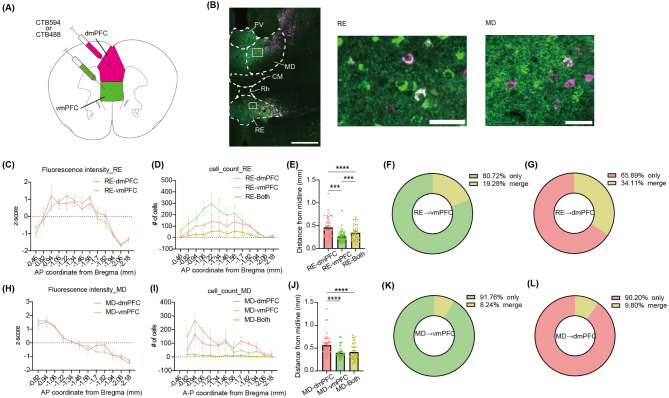
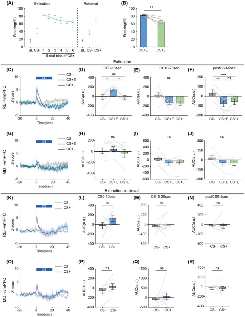
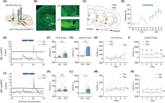
Animals adaptively regulate aversive memories in safe environments through extinction, a process central to exposure therapy for anxiety disorders. The limbic thalamus controls cognitive function in concert with interconnected cortical and limbic structures. Though medial prefrontal (mPFC) afferents to the limbic thalamus regulate aversive memory, the functional role of limbic thalamus efferents to mPFC is unclear. Here, we investigated the roles of thalamic nuclei, the reuniens (RE) and mediodorsal (MD) thalamus, projecting to the medial prefrontal cortex (mPFC) in aversive memory conditioning and extinction in male mice. Using retrograde tracing, we demonstrated that ventromedial PFC (vmPFC)- and dorsomedial PFC (dmPFC)-projecting neurons are topologically segregated within the RE and MD. Fiber photometry revealed that both RE→vmPFC and MD→vmPFC neurons respond to aversive stimuli. Notably, RE→vmPFC neurons develop shock-associated cue (CS+) response during aversive conditioning. During extinction, RE→vmPFC neurons exhibited a biphasic response to CS+, while MD→vmPFC neurons showed no cue-evoked activity. Neither optogenetic activation nor inactivation of these populations altered freezing behavior during extinction compared to controls. Collectively, these findings indicate that RE→vmPFC neurons encode aversive cue information during extinction but are dispensable for behavioral modulation. This study highlights the distinct contributions of limbic thalamus-PFC circuits to aversive memory processing.
期刊介绍:
Molecular Brain is an open access, peer-reviewed journal that considers manuscripts on all aspects of studies on the nervous system at the molecular, cellular, and systems level providing a forum for scientists to communicate their findings.
Molecular brain research is a rapidly expanding research field in which integrative approaches at the genetic, molecular, cellular and synaptic levels yield key information about the physiological and pathological brain. These studies involve the use of a wide range of modern techniques in molecular biology, genomics, proteomics, imaging and electrophysiology.
 求助内容:
求助内容: 应助结果提醒方式:


