Anna K Barton, Jacek Kwiecinski, Hidenobu Hashimoto, Mark Hyun, Keiichiro Kuronuma, Aditya Killekar, Aakriti Gupta, Nipun Manral, John Moore, Marc R Dweck, David E Newby, Daniel S Berman, Damini Dey, Piotr Slomka
{"title":"使用正电子发射断层扫描和计算机断层扫描成像小动态病变:18f -氟化钠瓣膜幻像研究","authors":"Anna K Barton, Jacek Kwiecinski, Hidenobu Hashimoto, Mark Hyun, Keiichiro Kuronuma, Aditya Killekar, Aakriti Gupta, Nipun Manral, John Moore, Marc R Dweck, David E Newby, Daniel S Berman, Damini Dey, Piotr Slomka","doi":"10.1093/ehjimp/qyaf013","DOIUrl":null,"url":null,"abstract":"<p><strong>Aims: </strong><sup>18</sup>F-sodium fluoride (<sup>18</sup>F-NaF) positron emission tomography (PET) detects active microcalcification and predicts adverse outcomes including bioprosthetic valve deterioration. However, measuring small areas of <sup>18</sup>F-NaF uptake within moving structures remains challenging, requiring further optimization. We developed a representative cardiac phantom to optimize <sup>18</sup>F-NaF imaging of bioprosthetic valves.</p><p><strong>Methods and results: </strong>We placed a bioprosthetic valve with two pockets sutured to the leaflets mimicking valvular lesions and a subvalvular ring mimicking the valve remnant into the phantom and injected each with <sup>18</sup>F-radionuclide (1 μCi pockets, 4 μCi ring). We injected the cardiac chambers with iohexol and <sup>18</sup>F-radionuclide (0.176 mCi) for background activity. PET and computed tomography (CT) images were acquired using a Siemens Biograph Vision high-resolution digital PET/CT scanner. We analysed target-to-background ratio (TBR) and signal-to-noise ratio (SNR) and subjective measures of image quality. We compared results with a human case of transcatheter aortic valve replacement. Initially the SNR and TBR in the phantom greatly exceeded those from human imaging. We reduced the scan duration used for reconstruction to 30 and 15 s, achieving comparable results (30 s vs. 15 s vs. patient: SNR 45.6 vs. 13.9 vs. 44.3, TBR<sub>max</sub> 6.5 vs. 5.4 vs. 4.1, noise 10.2% vs. 8.8% vs. 12.0%). With motion correction, SNR and image quality improved in the phantom (30 s 135.8 vs. 45.6, 15 s 32.9 vs. 13.9) but remained similar in the human case (47.3 vs. 44.3).</p><p><strong>Conclusion: </strong>A cardiac phantom can mimic clinical <sup>18</sup>F-NaF valve bioprosthesis imaging, providing an opportunity to explore acquisition, reconstruction, and post-processing of <sup>18</sup>F-NaF PET/CT for small mobile cardiac structures.</p>","PeriodicalId":94317,"journal":{"name":"European heart journal. Imaging methods and practice","volume":"3 1","pages":"qyaf013"},"PeriodicalIF":0.0000,"publicationDate":"2025-02-28","publicationTypes":"Journal Article","fieldsOfStudy":null,"isOpenAccess":false,"openAccessPdf":"https://www.ncbi.nlm.nih.gov/pmc/articles/PMC11879021/pdf/","citationCount":"0","resultStr":"{\"title\":\"Imaging small dynamic lesions using positron emission tomography and computed tomography: an <sup>18</sup>F-sodium fluoride valvular phantom study.\",\"authors\":\"Anna K Barton, Jacek Kwiecinski, Hidenobu Hashimoto, Mark Hyun, Keiichiro Kuronuma, Aditya Killekar, Aakriti Gupta, Nipun Manral, John Moore, Marc R Dweck, David E Newby, Daniel S Berman, Damini Dey, Piotr Slomka\",\"doi\":\"10.1093/ehjimp/qyaf013\",\"DOIUrl\":null,\"url\":null,\"abstract\":\"<p><strong>Aims: </strong><sup>18</sup>F-sodium fluoride (<sup>18</sup>F-NaF) positron emission tomography (PET) detects active microcalcification and predicts adverse outcomes including bioprosthetic valve deterioration. However, measuring small areas of <sup>18</sup>F-NaF uptake within moving structures remains challenging, requiring further optimization. We developed a representative cardiac phantom to optimize <sup>18</sup>F-NaF imaging of bioprosthetic valves.</p><p><strong>Methods and results: </strong>We placed a bioprosthetic valve with two pockets sutured to the leaflets mimicking valvular lesions and a subvalvular ring mimicking the valve remnant into the phantom and injected each with <sup>18</sup>F-radionuclide (1 μCi pockets, 4 μCi ring). We injected the cardiac chambers with iohexol and <sup>18</sup>F-radionuclide (0.176 mCi) for background activity. PET and computed tomography (CT) images were acquired using a Siemens Biograph Vision high-resolution digital PET/CT scanner. We analysed target-to-background ratio (TBR) and signal-to-noise ratio (SNR) and subjective measures of image quality. We compared results with a human case of transcatheter aortic valve replacement. Initially the SNR and TBR in the phantom greatly exceeded those from human imaging. We reduced the scan duration used for reconstruction to 30 and 15 s, achieving comparable results (30 s vs. 15 s vs. patient: SNR 45.6 vs. 13.9 vs. 44.3, TBR<sub>max</sub> 6.5 vs. 5.4 vs. 4.1, noise 10.2% vs. 8.8% vs. 12.0%). With motion correction, SNR and image quality improved in the phantom (30 s 135.8 vs. 45.6, 15 s 32.9 vs. 13.9) but remained similar in the human case (47.3 vs. 44.3).</p><p><strong>Conclusion: </strong>A cardiac phantom can mimic clinical <sup>18</sup>F-NaF valve bioprosthesis imaging, providing an opportunity to explore acquisition, reconstruction, and post-processing of <sup>18</sup>F-NaF PET/CT for small mobile cardiac structures.</p>\",\"PeriodicalId\":94317,\"journal\":{\"name\":\"European heart journal. Imaging methods and practice\",\"volume\":\"3 1\",\"pages\":\"qyaf013\"},\"PeriodicalIF\":0.0000,\"publicationDate\":\"2025-02-28\",\"publicationTypes\":\"Journal Article\",\"fieldsOfStudy\":null,\"isOpenAccess\":false,\"openAccessPdf\":\"https://www.ncbi.nlm.nih.gov/pmc/articles/PMC11879021/pdf/\",\"citationCount\":\"0\",\"resultStr\":null,\"platform\":\"Semanticscholar\",\"paperid\":null,\"PeriodicalName\":\"European heart journal. Imaging methods and practice\",\"FirstCategoryId\":\"1085\",\"ListUrlMain\":\"https://doi.org/10.1093/ehjimp/qyaf013\",\"RegionNum\":0,\"RegionCategory\":null,\"ArticlePicture\":[],\"TitleCN\":null,\"AbstractTextCN\":null,\"PMCID\":null,\"EPubDate\":\"2025/1/1 0:00:00\",\"PubModel\":\"eCollection\",\"JCR\":\"\",\"JCRName\":\"\",\"Score\":null,\"Total\":0}","platform":"Semanticscholar","paperid":null,"PeriodicalName":"European heart journal. Imaging methods and practice","FirstCategoryId":"1085","ListUrlMain":"https://doi.org/10.1093/ehjimp/qyaf013","RegionNum":0,"RegionCategory":null,"ArticlePicture":[],"TitleCN":null,"AbstractTextCN":null,"PMCID":null,"EPubDate":"2025/1/1 0:00:00","PubModel":"eCollection","JCR":"","JCRName":"","Score":null,"Total":0}
引用次数: 0
摘要
目的:18f -氟化钠(18F-NaF)正电子发射断层扫描(PET)检测活动性微钙化并预测包括生物瓣膜恶化在内的不良后果。然而,在移动结构中测量小区域的18F-NaF吸收仍然具有挑战性,需要进一步优化。我们开发了一个具有代表性的心脏幻影来优化生物假体瓣膜的18F-NaF成像。方法和结果:我们将一个生物假体瓣膜置入假体中,假体瓣膜有两个囊状结构,分别与模拟瓣膜病变的小叶和模拟瓣膜残余物的瓣下环缝合,并分别注射18f -核素(囊状结构1 μCi,环状结构4 μCi)。我们给心脏腔注射碘己醇和18f放射性核素(0.176 mCi)测定本底活性。使用西门子Biograph Vision高分辨率数字PET/CT扫描仪获取PET和CT图像。我们分析了目标与背景比(TBR)和信噪比(SNR)以及图像质量的主观度量。我们将结果与一例经导管主动脉瓣置换术进行比较。最初,幻影中的信噪比和TBR大大超过了人类成像。我们将用于重建的扫描时间缩短至30秒和15秒,获得了可比较的结果(30秒vs 15秒vs患者:信噪比45.6 vs 13.9 vs 44.3, TBRmax 6.5 vs 5.4 vs 4.1,噪声10.2% vs 8.8% vs 12.0%)。通过运动校正,幻影的信噪比和图像质量得到改善(30秒135.8比45.6,15秒32.9比13.9),但在人类病例中保持相似(47.3比44.3)。结论:心脏幻影可以模拟临床18F-NaF瓣膜生物假体成像,为探索18F-NaF PET/CT对小型移动心脏结构的采集、重建和后处理提供了机会。
Imaging small dynamic lesions using positron emission tomography and computed tomography: an 18F-sodium fluoride valvular phantom study.
Aims: 18F-sodium fluoride (18F-NaF) positron emission tomography (PET) detects active microcalcification and predicts adverse outcomes including bioprosthetic valve deterioration. However, measuring small areas of 18F-NaF uptake within moving structures remains challenging, requiring further optimization. We developed a representative cardiac phantom to optimize 18F-NaF imaging of bioprosthetic valves.
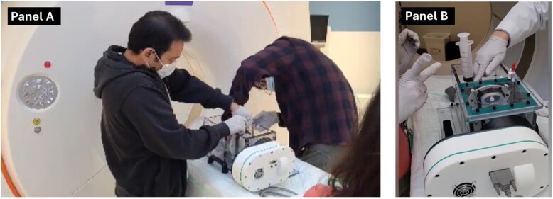
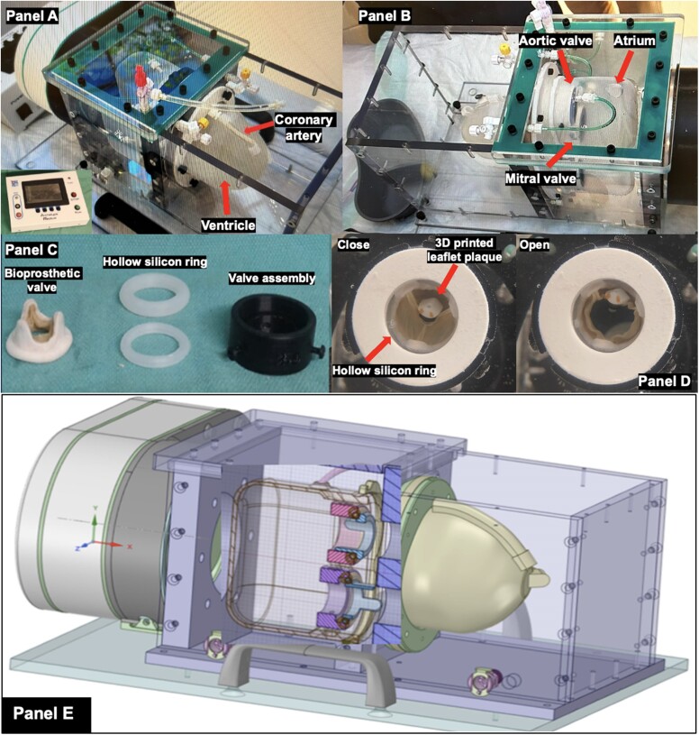
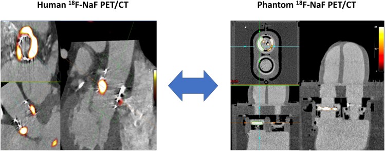
Methods and results: We placed a bioprosthetic valve with two pockets sutured to the leaflets mimicking valvular lesions and a subvalvular ring mimicking the valve remnant into the phantom and injected each with 18F-radionuclide (1 μCi pockets, 4 μCi ring). We injected the cardiac chambers with iohexol and 18F-radionuclide (0.176 mCi) for background activity. PET and computed tomography (CT) images were acquired using a Siemens Biograph Vision high-resolution digital PET/CT scanner. We analysed target-to-background ratio (TBR) and signal-to-noise ratio (SNR) and subjective measures of image quality. We compared results with a human case of transcatheter aortic valve replacement. Initially the SNR and TBR in the phantom greatly exceeded those from human imaging. We reduced the scan duration used for reconstruction to 30 and 15 s, achieving comparable results (30 s vs. 15 s vs. patient: SNR 45.6 vs. 13.9 vs. 44.3, TBRmax 6.5 vs. 5.4 vs. 4.1, noise 10.2% vs. 8.8% vs. 12.0%). With motion correction, SNR and image quality improved in the phantom (30 s 135.8 vs. 45.6, 15 s 32.9 vs. 13.9) but remained similar in the human case (47.3 vs. 44.3).
Conclusion: A cardiac phantom can mimic clinical 18F-NaF valve bioprosthesis imaging, providing an opportunity to explore acquisition, reconstruction, and post-processing of 18F-NaF PET/CT for small mobile cardiac structures.

 求助内容:
求助内容: 应助结果提醒方式:


