Kacper Żebrowski, Małgorzata Kandefer-Gola, Jolanta Bujok, Wojciech Borawski, Stanisław Dzimira
{"title":"犬输尿管血管肉瘤:罕见原发肿瘤伴不寻常的对侧肾脏转移。","authors":"Kacper Żebrowski, Małgorzata Kandefer-Gola, Jolanta Bujok, Wojciech Borawski, Stanisław Dzimira","doi":"10.1155/crve/3429069","DOIUrl":null,"url":null,"abstract":"<p><p>A dog (neutered male, 11 years old, Labrador retriever) underwent abdominal ultrasound, which revealed a 7 cm diameter tumour (caudal region of the left kidney). The animal showed symptoms of weight loss, apathy, haematuria, and abdominal pain. A computed tomography (CT) scan confirmed the presence of a tumour originating from the ureter. Following surgery to remove the ureter with the attached kidney, a histopathological examination was performed. The tumour was classified as a haemangiosarcoma. After the initial recovery, 2 months after surgery, the dog was diagnosed with a tumour in the other kidney. A fine needle biopsy was carried out. A haemangiosarcoma metastasis was suspected. Neoplasms of the ureter are a rare pathology. This is the first case in which metastasis to the second kidney has been confirmed.</p>","PeriodicalId":37339,"journal":{"name":"Case Reports in Veterinary Medicine","volume":"2025 ","pages":"3429069"},"PeriodicalIF":0.0000,"publicationDate":"2025-02-05","publicationTypes":"Journal Article","fieldsOfStudy":null,"isOpenAccess":false,"openAccessPdf":"https://www.ncbi.nlm.nih.gov/pmc/articles/PMC11870759/pdf/","citationCount":"0","resultStr":"{\"title\":\"Ureteral Haemangiosarcoma in a Dog: Rare Primary Tumour With Unusual Metastasis to the Contralateral Kidney.\",\"authors\":\"Kacper Żebrowski, Małgorzata Kandefer-Gola, Jolanta Bujok, Wojciech Borawski, Stanisław Dzimira\",\"doi\":\"10.1155/crve/3429069\",\"DOIUrl\":null,\"url\":null,\"abstract\":\"<p><p>A dog (neutered male, 11 years old, Labrador retriever) underwent abdominal ultrasound, which revealed a 7 cm diameter tumour (caudal region of the left kidney). The animal showed symptoms of weight loss, apathy, haematuria, and abdominal pain. A computed tomography (CT) scan confirmed the presence of a tumour originating from the ureter. Following surgery to remove the ureter with the attached kidney, a histopathological examination was performed. The tumour was classified as a haemangiosarcoma. After the initial recovery, 2 months after surgery, the dog was diagnosed with a tumour in the other kidney. A fine needle biopsy was carried out. A haemangiosarcoma metastasis was suspected. Neoplasms of the ureter are a rare pathology. This is the first case in which metastasis to the second kidney has been confirmed.</p>\",\"PeriodicalId\":37339,\"journal\":{\"name\":\"Case Reports in Veterinary Medicine\",\"volume\":\"2025 \",\"pages\":\"3429069\"},\"PeriodicalIF\":0.0000,\"publicationDate\":\"2025-02-05\",\"publicationTypes\":\"Journal Article\",\"fieldsOfStudy\":null,\"isOpenAccess\":false,\"openAccessPdf\":\"https://www.ncbi.nlm.nih.gov/pmc/articles/PMC11870759/pdf/\",\"citationCount\":\"0\",\"resultStr\":null,\"platform\":\"Semanticscholar\",\"paperid\":null,\"PeriodicalName\":\"Case Reports in Veterinary Medicine\",\"FirstCategoryId\":\"1085\",\"ListUrlMain\":\"https://doi.org/10.1155/crve/3429069\",\"RegionNum\":0,\"RegionCategory\":null,\"ArticlePicture\":[],\"TitleCN\":null,\"AbstractTextCN\":null,\"PMCID\":null,\"EPubDate\":\"2025/1/1 0:00:00\",\"PubModel\":\"eCollection\",\"JCR\":\"Q3\",\"JCRName\":\"Veterinary\",\"Score\":null,\"Total\":0}","platform":"Semanticscholar","paperid":null,"PeriodicalName":"Case Reports in Veterinary Medicine","FirstCategoryId":"1085","ListUrlMain":"https://doi.org/10.1155/crve/3429069","RegionNum":0,"RegionCategory":null,"ArticlePicture":[],"TitleCN":null,"AbstractTextCN":null,"PMCID":null,"EPubDate":"2025/1/1 0:00:00","PubModel":"eCollection","JCR":"Q3","JCRName":"Veterinary","Score":null,"Total":0}
Ureteral Haemangiosarcoma in a Dog: Rare Primary Tumour With Unusual Metastasis to the Contralateral Kidney.
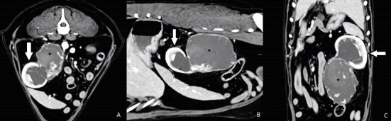
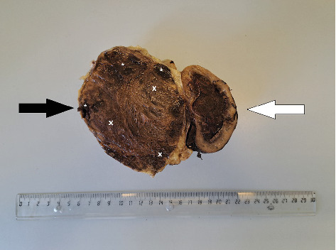
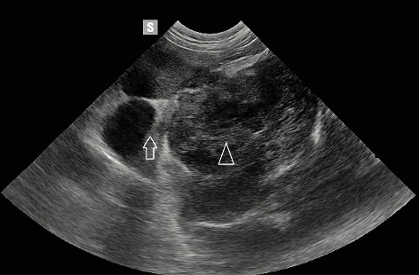
A dog (neutered male, 11 years old, Labrador retriever) underwent abdominal ultrasound, which revealed a 7 cm diameter tumour (caudal region of the left kidney). The animal showed symptoms of weight loss, apathy, haematuria, and abdominal pain. A computed tomography (CT) scan confirmed the presence of a tumour originating from the ureter. Following surgery to remove the ureter with the attached kidney, a histopathological examination was performed. The tumour was classified as a haemangiosarcoma. After the initial recovery, 2 months after surgery, the dog was diagnosed with a tumour in the other kidney. A fine needle biopsy was carried out. A haemangiosarcoma metastasis was suspected. Neoplasms of the ureter are a rare pathology. This is the first case in which metastasis to the second kidney has been confirmed.
期刊介绍:
Case Reports in Veterinary Medicine is a peer-reviewed, Open Access journal that publishes case reports and case series in all areas of veterinary medicine.

 求助内容:
求助内容: 应助结果提醒方式:


