{"title":"5-T磁共振成像显示人脑血管周围空间。","authors":"Sirui Liu, Jianbo Li, Rui Hua, Yaowen Xing, Jiaojiao Wu, Jiang Lin, Jian Wang, Yan Shan, Lei Xu, Feng Shi, Mengsu Zeng","doi":"10.1186/s12868-025-00925-z","DOIUrl":null,"url":null,"abstract":"<p><strong>Background: </strong>To evaluate the effectiveness of 5-Tesla (T) magnetic resonance imaging (MRI) in the visualization of perivascular spaces (PVS).</p><p><strong>Method: </strong>A total of seventeen subjects underwent three-dimensional (3D) T1- and T2-weighted imaging on both 3-T and 5-T MRI systems. Twelve of these subjects underwent quantitative analysis of PVS in the semioval center (SOC) and basal ganglia (BG), with comparisons made between the two systems using paired-sample Wilcoxon tests. Additionally, high-resolution 5-T images were acquired for five other participants to examine the detailed anatomy of PVS in the SOC, BG, and cerebral cortex.</p><p><strong>Results: </strong>Compared with 3-T MRI, 5-T MRI detected more PVS in the SOC and BG [39.5 (32.0-63.0) vs. 56.5 (44.0-75.5) and 49.5 (27.0-55.8) vs. 65.5 (53.0-72.0)] with p-values of 0.002 and 0.004, respectively. In these two regions, the PVS tortuosity, defined as the ratio of the actual path length to the straight-line distance between the start and end points of the PVS, was lower at 3-T compared to 5-T (p = 0.012 for the SOC and p = 0.006 for the BG). The length of PVS in the SOC on 5-T was longer than those on 3-T [4.6 mm (3.9-6.3 mm) vs. 5.1 mm (4.6-6.7 mm), p = 0.049]. In addition, the 5-T MRI provided enhanced visualization of the morphology of PVS in vivo, and improved the depiction of PVS across various brain regions, especially in the cortex, illustrating their course and associated small vessels.</p><p><strong>Conclusions: </strong>5-T MRI notably enhanced the visualization of PVS compared to 3-T, particularly in its ability to depict PVS anatomy in the cortex using high-resolution images. This advancement may pave the way for further research into the physiological roles of PVS and their involvement in related diseases.</p>","PeriodicalId":9031,"journal":{"name":"BMC Neuroscience","volume":"26 1","pages":"18"},"PeriodicalIF":2.3000,"publicationDate":"2025-03-03","publicationTypes":"Journal Article","fieldsOfStudy":null,"isOpenAccess":false,"openAccessPdf":"https://www.ncbi.nlm.nih.gov/pmc/articles/PMC11877699/pdf/","citationCount":"0","resultStr":"{\"title\":\"Visualization of perivascular spaces in the human brain with 5-T magnetic resonance imaging.\",\"authors\":\"Sirui Liu, Jianbo Li, Rui Hua, Yaowen Xing, Jiaojiao Wu, Jiang Lin, Jian Wang, Yan Shan, Lei Xu, Feng Shi, Mengsu Zeng\",\"doi\":\"10.1186/s12868-025-00925-z\",\"DOIUrl\":null,\"url\":null,\"abstract\":\"<p><strong>Background: </strong>To evaluate the effectiveness of 5-Tesla (T) magnetic resonance imaging (MRI) in the visualization of perivascular spaces (PVS).</p><p><strong>Method: </strong>A total of seventeen subjects underwent three-dimensional (3D) T1- and T2-weighted imaging on both 3-T and 5-T MRI systems. Twelve of these subjects underwent quantitative analysis of PVS in the semioval center (SOC) and basal ganglia (BG), with comparisons made between the two systems using paired-sample Wilcoxon tests. Additionally, high-resolution 5-T images were acquired for five other participants to examine the detailed anatomy of PVS in the SOC, BG, and cerebral cortex.</p><p><strong>Results: </strong>Compared with 3-T MRI, 5-T MRI detected more PVS in the SOC and BG [39.5 (32.0-63.0) vs. 56.5 (44.0-75.5) and 49.5 (27.0-55.8) vs. 65.5 (53.0-72.0)] with p-values of 0.002 and 0.004, respectively. In these two regions, the PVS tortuosity, defined as the ratio of the actual path length to the straight-line distance between the start and end points of the PVS, was lower at 3-T compared to 5-T (p = 0.012 for the SOC and p = 0.006 for the BG). The length of PVS in the SOC on 5-T was longer than those on 3-T [4.6 mm (3.9-6.3 mm) vs. 5.1 mm (4.6-6.7 mm), p = 0.049]. In addition, the 5-T MRI provided enhanced visualization of the morphology of PVS in vivo, and improved the depiction of PVS across various brain regions, especially in the cortex, illustrating their course and associated small vessels.</p><p><strong>Conclusions: </strong>5-T MRI notably enhanced the visualization of PVS compared to 3-T, particularly in its ability to depict PVS anatomy in the cortex using high-resolution images. This advancement may pave the way for further research into the physiological roles of PVS and their involvement in related diseases.</p>\",\"PeriodicalId\":9031,\"journal\":{\"name\":\"BMC Neuroscience\",\"volume\":\"26 1\",\"pages\":\"18\"},\"PeriodicalIF\":2.3000,\"publicationDate\":\"2025-03-03\",\"publicationTypes\":\"Journal Article\",\"fieldsOfStudy\":null,\"isOpenAccess\":false,\"openAccessPdf\":\"https://www.ncbi.nlm.nih.gov/pmc/articles/PMC11877699/pdf/\",\"citationCount\":\"0\",\"resultStr\":null,\"platform\":\"Semanticscholar\",\"paperid\":null,\"PeriodicalName\":\"BMC Neuroscience\",\"FirstCategoryId\":\"3\",\"ListUrlMain\":\"https://doi.org/10.1186/s12868-025-00925-z\",\"RegionNum\":4,\"RegionCategory\":\"医学\",\"ArticlePicture\":[],\"TitleCN\":null,\"AbstractTextCN\":null,\"PMCID\":null,\"EPubDate\":\"\",\"PubModel\":\"\",\"JCR\":\"Q3\",\"JCRName\":\"NEUROSCIENCES\",\"Score\":null,\"Total\":0}","platform":"Semanticscholar","paperid":null,"PeriodicalName":"BMC Neuroscience","FirstCategoryId":"3","ListUrlMain":"https://doi.org/10.1186/s12868-025-00925-z","RegionNum":4,"RegionCategory":"医学","ArticlePicture":[],"TitleCN":null,"AbstractTextCN":null,"PMCID":null,"EPubDate":"","PubModel":"","JCR":"Q3","JCRName":"NEUROSCIENCES","Score":null,"Total":0}
引用次数: 0
摘要
背景:评价5-特斯拉(T)磁共振成像(MRI)在血管周围间隙(PVS)显示中的有效性。方法:共17例受试者在3-T和5-T MRI系统上进行了三维(3D) T1和t2加权成像。其中12名受试者在半动中枢(SOC)和基底神经节(BG)进行了PVS的定量分析,并使用配对样本Wilcoxon测试对两个系统进行了比较。此外,对另外五名参与者获取高分辨率5-T图像,以检查SOC, BG和大脑皮层的PVS详细解剖结构。结果:与3-T MRI相比,5-T MRI在SOC和BG中检测到更多的PVS [39.5 (32.0-63.0) vs. 56.5(44.0-75.5)和49.5 (27.0-55.8)vs. 65.5 (53.0-72.0)], p值分别为0.002和0.004。在这两个区域,与5-T相比,3-T的PVS扭曲度(定义为实际路径长度与PVS起点和终点之间的直线距离之比)较低(SOC为p = 0.012, BG为p = 0.006)。5-T上SOC中PVS的长度比3-T上更长[4.6 mm (3.9-6.3 mm)比5.1 mm (4.6-6.7 mm), p = 0.049]。此外,5-T MRI增强了PVS在体内形态的可视化,并改善了PVS在大脑各区域,特别是皮层的描述,显示了它们的路线和相关的小血管。结论:与3-T相比,5-T MRI显着增强了PVS的可视化,特别是在使用高分辨率图像描绘皮层PVS解剖的能力方面。这一进展可能为进一步研究PVS的生理作用及其在相关疾病中的作用铺平道路。
Visualization of perivascular spaces in the human brain with 5-T magnetic resonance imaging.
Background: To evaluate the effectiveness of 5-Tesla (T) magnetic resonance imaging (MRI) in the visualization of perivascular spaces (PVS).
Method: A total of seventeen subjects underwent three-dimensional (3D) T1- and T2-weighted imaging on both 3-T and 5-T MRI systems. Twelve of these subjects underwent quantitative analysis of PVS in the semioval center (SOC) and basal ganglia (BG), with comparisons made between the two systems using paired-sample Wilcoxon tests. Additionally, high-resolution 5-T images were acquired for five other participants to examine the detailed anatomy of PVS in the SOC, BG, and cerebral cortex.
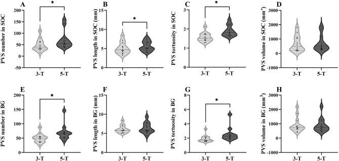
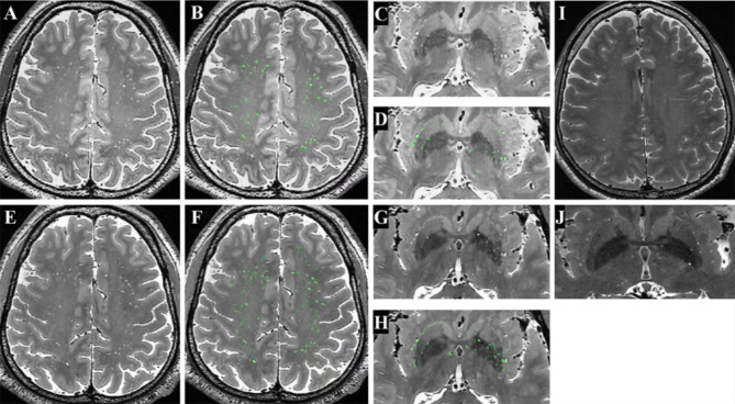
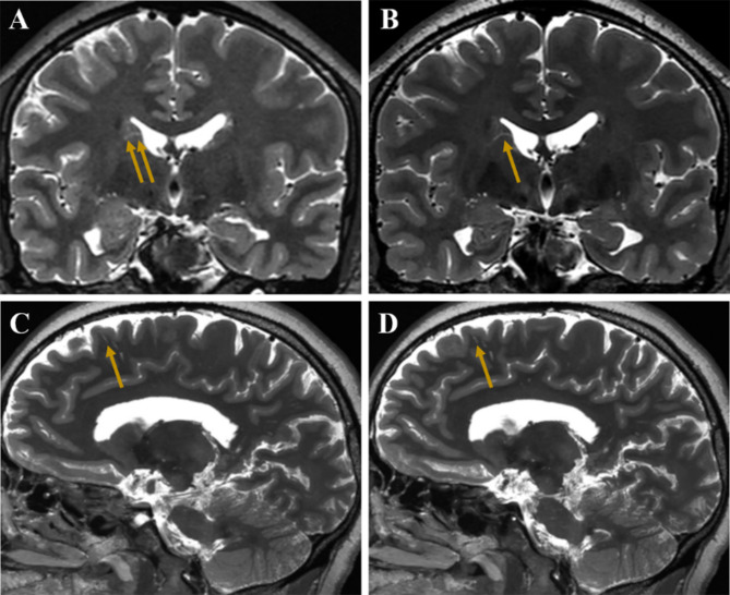
Results: Compared with 3-T MRI, 5-T MRI detected more PVS in the SOC and BG [39.5 (32.0-63.0) vs. 56.5 (44.0-75.5) and 49.5 (27.0-55.8) vs. 65.5 (53.0-72.0)] with p-values of 0.002 and 0.004, respectively. In these two regions, the PVS tortuosity, defined as the ratio of the actual path length to the straight-line distance between the start and end points of the PVS, was lower at 3-T compared to 5-T (p = 0.012 for the SOC and p = 0.006 for the BG). The length of PVS in the SOC on 5-T was longer than those on 3-T [4.6 mm (3.9-6.3 mm) vs. 5.1 mm (4.6-6.7 mm), p = 0.049]. In addition, the 5-T MRI provided enhanced visualization of the morphology of PVS in vivo, and improved the depiction of PVS across various brain regions, especially in the cortex, illustrating their course and associated small vessels.
Conclusions: 5-T MRI notably enhanced the visualization of PVS compared to 3-T, particularly in its ability to depict PVS anatomy in the cortex using high-resolution images. This advancement may pave the way for further research into the physiological roles of PVS and their involvement in related diseases.
期刊介绍:
BMC Neuroscience is an open access, peer-reviewed journal that considers articles on all aspects of neuroscience, welcoming studies that provide insight into the molecular, cellular, developmental, genetic and genomic, systems, network, cognitive and behavioral aspects of nervous system function in both health and disease. Both experimental and theoretical studies are within scope, as are studies that describe methodological approaches to monitoring or manipulating nervous system function.
 求助内容:
求助内容: 应助结果提醒方式:


