Jueun Sim, Chan E. Park, In Cho, Kyeongbae Min, Minho Eom, Seungjae Han, Hyungju Jeon, Eun-Seo Cho, Yunjeong Lee, Young Hyun Yun, Sungho Lee, Deok-Hyeon Cheon, Jihyun Kim, Museong Kim, Hyun-Ju Cho, Ji-Won Park, Ajeet Kumar, Yosep Chong, Jeong Seuk Kang, Kiryl D. Piatkevich, Erica E. Jung, Du-Seock Kang, Seok-Kyu Kwon, Jinhyun Kim, Ki-Jun Yoon, Jeong-Soo Lee, Cheol-Hee Kim, Myunghwan Choi, Jin Woo Kim, Mi-Ryoung Song, Hyung Jin Choi, Edward S. Boyden, Young-Gyu Yoon* and Jae-Byum Chang*,
{"title":"利用扩增显微镜对整个小鼠胚胎进行纳米级分辨率成像","authors":"Jueun Sim, Chan E. Park, In Cho, Kyeongbae Min, Minho Eom, Seungjae Han, Hyungju Jeon, Eun-Seo Cho, Yunjeong Lee, Young Hyun Yun, Sungho Lee, Deok-Hyeon Cheon, Jihyun Kim, Museong Kim, Hyun-Ju Cho, Ji-Won Park, Ajeet Kumar, Yosep Chong, Jeong Seuk Kang, Kiryl D. Piatkevich, Erica E. Jung, Du-Seock Kang, Seok-Kyu Kwon, Jinhyun Kim, Ki-Jun Yoon, Jeong-Soo Lee, Cheol-Hee Kim, Myunghwan Choi, Jin Woo Kim, Mi-Ryoung Song, Hyung Jin Choi, Edward S. Boyden, Young-Gyu Yoon* and Jae-Byum Chang*, ","doi":"10.1021/acsnano.4c1479110.1021/acsnano.4c14791","DOIUrl":null,"url":null,"abstract":"<p >Nanoscale imaging of whole vertebrates is essential for the systematic understanding of human diseases, yet this goal has not yet been achieved. Expansion microscopy (ExM) is an attractive option for accomplishing this aim; however, the expansion of even mouse embryos at mid- and late-developmental stages, which have fewer calcified body parts than adult mice, is yet to be demonstrated due to the challenges of expanding calcified tissues. Here, we introduce a state-of-the-art ExM technique, termed whole-body ExM, that utilizes cyclic digestion. This technique allows for the super-resolution, volumetric imaging of anatomical structures, proteins, and endogenous fluorescent proteins (FPs) within embryonic and neonatal mice by expanding them 4-fold. The key feature of whole-body ExM is the alternating application of two enzyme compositions repeated multiple times. Through the simple repetition of this digestion process with an increasing number of cycles, mouse embryos of various stages up to E18.5, and even neonatal mice, which display a dramatic difference in the content of calcified tissues compared to embryos, are expanded without further laborious optimization. Furthermore, the whole-body ExM’s ability to retain FP signals allows the visualization of various neuronal structures in transgenic mice. Whole-body ExM could facilitate studies of molecular changes in various vertebrates.</p>","PeriodicalId":21,"journal":{"name":"ACS Nano","volume":"19 8","pages":"7910–7927 7910–7927"},"PeriodicalIF":16.0000,"publicationDate":"2025-02-18","publicationTypes":"Journal Article","fieldsOfStudy":null,"isOpenAccess":false,"openAccessPdf":"","citationCount":"0","resultStr":"{\"title\":\"Nanoscale Resolution Imaging of Whole Mouse Embryos Using Expansion Microscopy\",\"authors\":\"Jueun Sim, Chan E. Park, In Cho, Kyeongbae Min, Minho Eom, Seungjae Han, Hyungju Jeon, Eun-Seo Cho, Yunjeong Lee, Young Hyun Yun, Sungho Lee, Deok-Hyeon Cheon, Jihyun Kim, Museong Kim, Hyun-Ju Cho, Ji-Won Park, Ajeet Kumar, Yosep Chong, Jeong Seuk Kang, Kiryl D. Piatkevich, Erica E. Jung, Du-Seock Kang, Seok-Kyu Kwon, Jinhyun Kim, Ki-Jun Yoon, Jeong-Soo Lee, Cheol-Hee Kim, Myunghwan Choi, Jin Woo Kim, Mi-Ryoung Song, Hyung Jin Choi, Edward S. Boyden, Young-Gyu Yoon* and Jae-Byum Chang*, \",\"doi\":\"10.1021/acsnano.4c1479110.1021/acsnano.4c14791\",\"DOIUrl\":null,\"url\":null,\"abstract\":\"<p >Nanoscale imaging of whole vertebrates is essential for the systematic understanding of human diseases, yet this goal has not yet been achieved. Expansion microscopy (ExM) is an attractive option for accomplishing this aim; however, the expansion of even mouse embryos at mid- and late-developmental stages, which have fewer calcified body parts than adult mice, is yet to be demonstrated due to the challenges of expanding calcified tissues. Here, we introduce a state-of-the-art ExM technique, termed whole-body ExM, that utilizes cyclic digestion. This technique allows for the super-resolution, volumetric imaging of anatomical structures, proteins, and endogenous fluorescent proteins (FPs) within embryonic and neonatal mice by expanding them 4-fold. The key feature of whole-body ExM is the alternating application of two enzyme compositions repeated multiple times. Through the simple repetition of this digestion process with an increasing number of cycles, mouse embryos of various stages up to E18.5, and even neonatal mice, which display a dramatic difference in the content of calcified tissues compared to embryos, are expanded without further laborious optimization. Furthermore, the whole-body ExM’s ability to retain FP signals allows the visualization of various neuronal structures in transgenic mice. Whole-body ExM could facilitate studies of molecular changes in various vertebrates.</p>\",\"PeriodicalId\":21,\"journal\":{\"name\":\"ACS Nano\",\"volume\":\"19 8\",\"pages\":\"7910–7927 7910–7927\"},\"PeriodicalIF\":16.0000,\"publicationDate\":\"2025-02-18\",\"publicationTypes\":\"Journal Article\",\"fieldsOfStudy\":null,\"isOpenAccess\":false,\"openAccessPdf\":\"\",\"citationCount\":\"0\",\"resultStr\":null,\"platform\":\"Semanticscholar\",\"paperid\":null,\"PeriodicalName\":\"ACS Nano\",\"FirstCategoryId\":\"88\",\"ListUrlMain\":\"https://pubs.acs.org/doi/10.1021/acsnano.4c14791\",\"RegionNum\":1,\"RegionCategory\":\"材料科学\",\"ArticlePicture\":[],\"TitleCN\":null,\"AbstractTextCN\":null,\"PMCID\":null,\"EPubDate\":\"\",\"PubModel\":\"\",\"JCR\":\"Q1\",\"JCRName\":\"CHEMISTRY, MULTIDISCIPLINARY\",\"Score\":null,\"Total\":0}","platform":"Semanticscholar","paperid":null,"PeriodicalName":"ACS Nano","FirstCategoryId":"88","ListUrlMain":"https://pubs.acs.org/doi/10.1021/acsnano.4c14791","RegionNum":1,"RegionCategory":"材料科学","ArticlePicture":[],"TitleCN":null,"AbstractTextCN":null,"PMCID":null,"EPubDate":"","PubModel":"","JCR":"Q1","JCRName":"CHEMISTRY, MULTIDISCIPLINARY","Score":null,"Total":0}
Nanoscale Resolution Imaging of Whole Mouse Embryos Using Expansion Microscopy
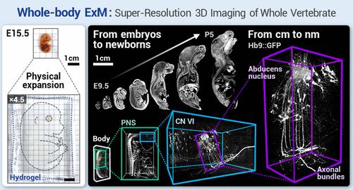
Nanoscale imaging of whole vertebrates is essential for the systematic understanding of human diseases, yet this goal has not yet been achieved. Expansion microscopy (ExM) is an attractive option for accomplishing this aim; however, the expansion of even mouse embryos at mid- and late-developmental stages, which have fewer calcified body parts than adult mice, is yet to be demonstrated due to the challenges of expanding calcified tissues. Here, we introduce a state-of-the-art ExM technique, termed whole-body ExM, that utilizes cyclic digestion. This technique allows for the super-resolution, volumetric imaging of anatomical structures, proteins, and endogenous fluorescent proteins (FPs) within embryonic and neonatal mice by expanding them 4-fold. The key feature of whole-body ExM is the alternating application of two enzyme compositions repeated multiple times. Through the simple repetition of this digestion process with an increasing number of cycles, mouse embryos of various stages up to E18.5, and even neonatal mice, which display a dramatic difference in the content of calcified tissues compared to embryos, are expanded without further laborious optimization. Furthermore, the whole-body ExM’s ability to retain FP signals allows the visualization of various neuronal structures in transgenic mice. Whole-body ExM could facilitate studies of molecular changes in various vertebrates.
期刊介绍:
ACS Nano, published monthly, serves as an international forum for comprehensive articles on nanoscience and nanotechnology research at the intersections of chemistry, biology, materials science, physics, and engineering. The journal fosters communication among scientists in these communities, facilitating collaboration, new research opportunities, and advancements through discoveries. ACS Nano covers synthesis, assembly, characterization, theory, and simulation of nanostructures, nanobiotechnology, nanofabrication, methods and tools for nanoscience and nanotechnology, and self- and directed-assembly. Alongside original research articles, it offers thorough reviews, perspectives on cutting-edge research, and discussions envisioning the future of nanoscience and nanotechnology.
 求助内容:
求助内容: 应助结果提醒方式:


