Maximilian Friedrich, Hyeck-Soo Son, Jasper Lind, Maximilian Hammer, Lizaveta Chychko, Timur Mert Yildirim, Gerd Uwe Auffarth, Victor Aristide Augustin
{"title":"术前水肿严重程度影响富克斯内皮性角膜营养不良患者行Descemet膜内皮角膜移植术后的预后:一项队列研究。","authors":"Maximilian Friedrich, Hyeck-Soo Son, Jasper Lind, Maximilian Hammer, Lizaveta Chychko, Timur Mert Yildirim, Gerd Uwe Auffarth, Victor Aristide Augustin","doi":"10.1186/s40662-025-00425-5","DOIUrl":null,"url":null,"abstract":"<p><strong>Background: </strong>In patients with Fuchs endothelial corneal dystrophy (FECD), the most beneficial stage to perform Descemet membrane endothelial keratoplasty (DMEK) remains uncertain. The goal of this study was to compare the surgical outcomes after DMEK in FECD patients with subclinical corneal edema and clinical corneal edema to test the hypothesis of whether performing surgery in subclinical corneal edema stages achieves better surgical outcomes.</p><p><strong>Methods: </strong>In this prospective, observational, single-institution cohort study, 106 pseudophakic eyes of 85 patients with FECD were divided into two groups depending on the presence of preoperative subclinical and clinical corneal edema. Subclinical corneal edema was diagnosed if more than one of the following criteria was present in Scheimpflug tomography: loss of regular isopachs, displacement of the thinnest point of the cornea, and focal posterior corneal surface depression. Clinical corneal edema was diagnosed with slit-lamp biomicroscopy. The primary outcome was the corrected distance visual acuity (CDVA) 4 months after DMEK. Secondary outcomes were central corneal thickness (CCT), thinnest corneal thickness (TCT), and total corneal density (TCD) in Scheimpflug tomography, as well as endothelial cell loss (ECL) and the re-bubbling rate. The differences between both groups were analyzed using clustered Wilcoxon rank-sum tests or a Chi-squared test.</p><p><strong>Results: </strong>Postoperative CDVA was significantly better in the group with subclinical edema (0.18 ± 0.12 logMAR) compared to the group with clinical edema (0.24 ± 0.19 logMAR; P = 0.026). Four months after DMEK, TCD was higher in the group with preoperative clinical edema [31.7 ± 8.3 gray scale units (GSU)] compared to the group with subclinical edema (27.8 ± 6.1 GSU; P = 0.005). The postoperative CCT, TCT, ECL, and re-bubbling rates did not differ significantly between both groups (all P > 0.05).</p><p><strong>Conclusions: </strong>DMEK for FECD yielded better visual acuity after 4 months when performed in the early stage of FECD compared to a later stage with clinical edema. This may be attributable to persistent corneal fibrosis after DMEK in eyes with preoperative clinically evident corneal edema, as suggested by higher postoperative corneal density in eyes with clinical edema. Consequently, the findings advocate for the consideration of earlier DMEK in FECD patients to achieve better surgical recovery.</p>","PeriodicalId":12194,"journal":{"name":"Eye and Vision","volume":"12 1","pages":"9"},"PeriodicalIF":4.0000,"publicationDate":"2025-03-01","publicationTypes":"Journal Article","fieldsOfStudy":null,"isOpenAccess":false,"openAccessPdf":"https://www.ncbi.nlm.nih.gov/pmc/articles/PMC11871603/pdf/","citationCount":"0","resultStr":"{\"title\":\"Preoperative edema severity affects outcomes after Descemet membrane endothelial keratoplasty for Fuchs endothelial corneal dystrophy: a cohort study.\",\"authors\":\"Maximilian Friedrich, Hyeck-Soo Son, Jasper Lind, Maximilian Hammer, Lizaveta Chychko, Timur Mert Yildirim, Gerd Uwe Auffarth, Victor Aristide Augustin\",\"doi\":\"10.1186/s40662-025-00425-5\",\"DOIUrl\":null,\"url\":null,\"abstract\":\"<p><strong>Background: </strong>In patients with Fuchs endothelial corneal dystrophy (FECD), the most beneficial stage to perform Descemet membrane endothelial keratoplasty (DMEK) remains uncertain. The goal of this study was to compare the surgical outcomes after DMEK in FECD patients with subclinical corneal edema and clinical corneal edema to test the hypothesis of whether performing surgery in subclinical corneal edema stages achieves better surgical outcomes.</p><p><strong>Methods: </strong>In this prospective, observational, single-institution cohort study, 106 pseudophakic eyes of 85 patients with FECD were divided into two groups depending on the presence of preoperative subclinical and clinical corneal edema. Subclinical corneal edema was diagnosed if more than one of the following criteria was present in Scheimpflug tomography: loss of regular isopachs, displacement of the thinnest point of the cornea, and focal posterior corneal surface depression. Clinical corneal edema was diagnosed with slit-lamp biomicroscopy. The primary outcome was the corrected distance visual acuity (CDVA) 4 months after DMEK. Secondary outcomes were central corneal thickness (CCT), thinnest corneal thickness (TCT), and total corneal density (TCD) in Scheimpflug tomography, as well as endothelial cell loss (ECL) and the re-bubbling rate. The differences between both groups were analyzed using clustered Wilcoxon rank-sum tests or a Chi-squared test.</p><p><strong>Results: </strong>Postoperative CDVA was significantly better in the group with subclinical edema (0.18 ± 0.12 logMAR) compared to the group with clinical edema (0.24 ± 0.19 logMAR; P = 0.026). Four months after DMEK, TCD was higher in the group with preoperative clinical edema [31.7 ± 8.3 gray scale units (GSU)] compared to the group with subclinical edema (27.8 ± 6.1 GSU; P = 0.005). The postoperative CCT, TCT, ECL, and re-bubbling rates did not differ significantly between both groups (all P > 0.05).</p><p><strong>Conclusions: </strong>DMEK for FECD yielded better visual acuity after 4 months when performed in the early stage of FECD compared to a later stage with clinical edema. This may be attributable to persistent corneal fibrosis after DMEK in eyes with preoperative clinically evident corneal edema, as suggested by higher postoperative corneal density in eyes with clinical edema. Consequently, the findings advocate for the consideration of earlier DMEK in FECD patients to achieve better surgical recovery.</p>\",\"PeriodicalId\":12194,\"journal\":{\"name\":\"Eye and Vision\",\"volume\":\"12 1\",\"pages\":\"9\"},\"PeriodicalIF\":4.0000,\"publicationDate\":\"2025-03-01\",\"publicationTypes\":\"Journal Article\",\"fieldsOfStudy\":null,\"isOpenAccess\":false,\"openAccessPdf\":\"https://www.ncbi.nlm.nih.gov/pmc/articles/PMC11871603/pdf/\",\"citationCount\":\"0\",\"resultStr\":null,\"platform\":\"Semanticscholar\",\"paperid\":null,\"PeriodicalName\":\"Eye and Vision\",\"FirstCategoryId\":\"3\",\"ListUrlMain\":\"https://doi.org/10.1186/s40662-025-00425-5\",\"RegionNum\":1,\"RegionCategory\":\"医学\",\"ArticlePicture\":[],\"TitleCN\":null,\"AbstractTextCN\":null,\"PMCID\":null,\"EPubDate\":\"\",\"PubModel\":\"\",\"JCR\":\"Q1\",\"JCRName\":\"OPHTHALMOLOGY\",\"Score\":null,\"Total\":0}","platform":"Semanticscholar","paperid":null,"PeriodicalName":"Eye and Vision","FirstCategoryId":"3","ListUrlMain":"https://doi.org/10.1186/s40662-025-00425-5","RegionNum":1,"RegionCategory":"医学","ArticlePicture":[],"TitleCN":null,"AbstractTextCN":null,"PMCID":null,"EPubDate":"","PubModel":"","JCR":"Q1","JCRName":"OPHTHALMOLOGY","Score":null,"Total":0}
Preoperative edema severity affects outcomes after Descemet membrane endothelial keratoplasty for Fuchs endothelial corneal dystrophy: a cohort study.
Background: In patients with Fuchs endothelial corneal dystrophy (FECD), the most beneficial stage to perform Descemet membrane endothelial keratoplasty (DMEK) remains uncertain. The goal of this study was to compare the surgical outcomes after DMEK in FECD patients with subclinical corneal edema and clinical corneal edema to test the hypothesis of whether performing surgery in subclinical corneal edema stages achieves better surgical outcomes.
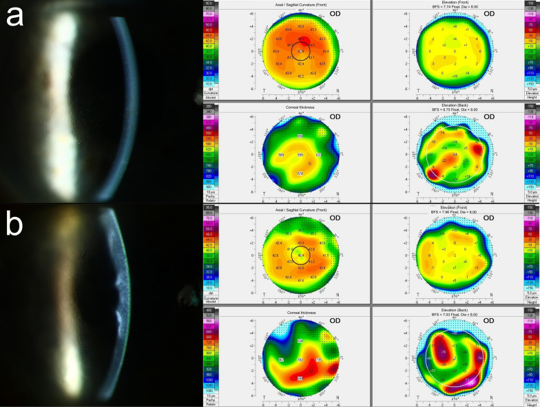
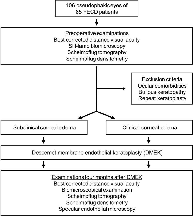
Methods: In this prospective, observational, single-institution cohort study, 106 pseudophakic eyes of 85 patients with FECD were divided into two groups depending on the presence of preoperative subclinical and clinical corneal edema. Subclinical corneal edema was diagnosed if more than one of the following criteria was present in Scheimpflug tomography: loss of regular isopachs, displacement of the thinnest point of the cornea, and focal posterior corneal surface depression. Clinical corneal edema was diagnosed with slit-lamp biomicroscopy. The primary outcome was the corrected distance visual acuity (CDVA) 4 months after DMEK. Secondary outcomes were central corneal thickness (CCT), thinnest corneal thickness (TCT), and total corneal density (TCD) in Scheimpflug tomography, as well as endothelial cell loss (ECL) and the re-bubbling rate. The differences between both groups were analyzed using clustered Wilcoxon rank-sum tests or a Chi-squared test.
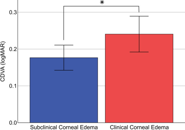
Results: Postoperative CDVA was significantly better in the group with subclinical edema (0.18 ± 0.12 logMAR) compared to the group with clinical edema (0.24 ± 0.19 logMAR; P = 0.026). Four months after DMEK, TCD was higher in the group with preoperative clinical edema [31.7 ± 8.3 gray scale units (GSU)] compared to the group with subclinical edema (27.8 ± 6.1 GSU; P = 0.005). The postoperative CCT, TCT, ECL, and re-bubbling rates did not differ significantly between both groups (all P > 0.05).
Conclusions: DMEK for FECD yielded better visual acuity after 4 months when performed in the early stage of FECD compared to a later stage with clinical edema. This may be attributable to persistent corneal fibrosis after DMEK in eyes with preoperative clinically evident corneal edema, as suggested by higher postoperative corneal density in eyes with clinical edema. Consequently, the findings advocate for the consideration of earlier DMEK in FECD patients to achieve better surgical recovery.
期刊介绍:
Eye and Vision is an open access, peer-reviewed journal for ophthalmologists and visual science specialists. It welcomes research articles, reviews, methodologies, commentaries, case reports, perspectives and short reports encompassing all aspects of eye and vision. Topics of interest include but are not limited to: current developments of theoretical, experimental and clinical investigations in ophthalmology, optometry and vision science which focus on novel and high-impact findings on central issues pertaining to biology, pathophysiology and etiology of eye diseases as well as advances in diagnostic techniques, surgical treatment, instrument updates, the latest drug findings, results of clinical trials and research findings. It aims to provide ophthalmologists and visual science specialists with the latest developments in theoretical, experimental and clinical investigations in eye and vision.
 求助内容:
求助内容: 应助结果提醒方式:


