Pengfei Jin, Linghui Zhang, Hong Yang, Tingting Jiang, Chenyang Xu, Jiehui Huang, Zhongyu Zhang, Lei Shi, Xu Wang
{"title":"改进的多参数CT诊断肾小实性肿块透明细胞癌的算法。","authors":"Pengfei Jin, Linghui Zhang, Hong Yang, Tingting Jiang, Chenyang Xu, Jiehui Huang, Zhongyu Zhang, Lei Shi, Xu Wang","doi":"10.1186/s40644-025-00847-3","DOIUrl":null,"url":null,"abstract":"<p><strong>Objective: </strong>To refine the existing CT algorithm to enhance inter-reader agreement and improve the diagnostic performance for clear-cell renal cell carcinoma (ccRCC) in solid renal masses less than 4 cm.</p><p><strong>Methods: </strong>A retrospective collection of 331 patients with pathologically confirmed renal masses were enrolled in this study. Two radiologists independently assessed the CT images: in addition to heterogeneity score (HS) and mass-to-cortex corticomedullary attenuation ratio (MCAR), measured parameters included ratio of major diameter to minor diameter at the maximum axial section (Major axis / Minor axis), tumor-renal interface, standardized heterogeneity ratio (SHR), and standardized nephrographic reduction rate (SNRR). Spearman's correlation analysis was performed to evaluate the relationship between SHR and HS. Univariate and multivariate logistic regression analyses were employed to identify independent risk factors and then CT-score was adjusted by those indicators. The diagnostic efficacy of the modified CT-scores was evaluated using ROC curve analysis.</p><p><strong>Results: </strong>The SHR and heterogeneity grade (HG) of mass were correlated positively with the HS (R = 0.749, 0.730, all P < 0.001). Logistic regression analysis determined that the Major axis / Minor axis (> 1.16), the tumor-renal interface (> 22.3 mm), and the SNRR (> 0.16) as additional independent risk factors to combine with HS and MCAR. Compared to the original CT-score, the two CT algorithms combined tumor-renal interface and SNRR showed significantly improved diagnostic efficacy for ccRCC (AUC: 0.770 vs. 0.861 and 0.862, all P < 0.001). The inter-observer agreement for HG was higher than that for HS (weighted Kappa coefficient: 0.797 vs. 0.722). The consistency of modified CT-score was also superior to original CT-score (weighted Kappa coefficient: 0.935 vs. 0.878).</p><p><strong>Conclusion: </strong>The modified CT algorithms not only enhanced inter-reader consistency but also improved the diagnostic capability for ccRCC in small renal masses.</p>","PeriodicalId":9548,"journal":{"name":"Cancer Imaging","volume":"25 1","pages":"22"},"PeriodicalIF":3.5000,"publicationDate":"2025-02-28","publicationTypes":"Journal Article","fieldsOfStudy":null,"isOpenAccess":false,"openAccessPdf":"https://www.ncbi.nlm.nih.gov/pmc/articles/PMC11869432/pdf/","citationCount":"0","resultStr":"{\"title\":\"Development of modified multi-parametric CT algorithms for diagnosing clear-cell renal cell carcinoma in small solid renal masses.\",\"authors\":\"Pengfei Jin, Linghui Zhang, Hong Yang, Tingting Jiang, Chenyang Xu, Jiehui Huang, Zhongyu Zhang, Lei Shi, Xu Wang\",\"doi\":\"10.1186/s40644-025-00847-3\",\"DOIUrl\":null,\"url\":null,\"abstract\":\"<p><strong>Objective: </strong>To refine the existing CT algorithm to enhance inter-reader agreement and improve the diagnostic performance for clear-cell renal cell carcinoma (ccRCC) in solid renal masses less than 4 cm.</p><p><strong>Methods: </strong>A retrospective collection of 331 patients with pathologically confirmed renal masses were enrolled in this study. Two radiologists independently assessed the CT images: in addition to heterogeneity score (HS) and mass-to-cortex corticomedullary attenuation ratio (MCAR), measured parameters included ratio of major diameter to minor diameter at the maximum axial section (Major axis / Minor axis), tumor-renal interface, standardized heterogeneity ratio (SHR), and standardized nephrographic reduction rate (SNRR). Spearman's correlation analysis was performed to evaluate the relationship between SHR and HS. Univariate and multivariate logistic regression analyses were employed to identify independent risk factors and then CT-score was adjusted by those indicators. The diagnostic efficacy of the modified CT-scores was evaluated using ROC curve analysis.</p><p><strong>Results: </strong>The SHR and heterogeneity grade (HG) of mass were correlated positively with the HS (R = 0.749, 0.730, all P < 0.001). Logistic regression analysis determined that the Major axis / Minor axis (> 1.16), the tumor-renal interface (> 22.3 mm), and the SNRR (> 0.16) as additional independent risk factors to combine with HS and MCAR. Compared to the original CT-score, the two CT algorithms combined tumor-renal interface and SNRR showed significantly improved diagnostic efficacy for ccRCC (AUC: 0.770 vs. 0.861 and 0.862, all P < 0.001). The inter-observer agreement for HG was higher than that for HS (weighted Kappa coefficient: 0.797 vs. 0.722). The consistency of modified CT-score was also superior to original CT-score (weighted Kappa coefficient: 0.935 vs. 0.878).</p><p><strong>Conclusion: </strong>The modified CT algorithms not only enhanced inter-reader consistency but also improved the diagnostic capability for ccRCC in small renal masses.</p>\",\"PeriodicalId\":9548,\"journal\":{\"name\":\"Cancer Imaging\",\"volume\":\"25 1\",\"pages\":\"22\"},\"PeriodicalIF\":3.5000,\"publicationDate\":\"2025-02-28\",\"publicationTypes\":\"Journal Article\",\"fieldsOfStudy\":null,\"isOpenAccess\":false,\"openAccessPdf\":\"https://www.ncbi.nlm.nih.gov/pmc/articles/PMC11869432/pdf/\",\"citationCount\":\"0\",\"resultStr\":null,\"platform\":\"Semanticscholar\",\"paperid\":null,\"PeriodicalName\":\"Cancer Imaging\",\"FirstCategoryId\":\"3\",\"ListUrlMain\":\"https://doi.org/10.1186/s40644-025-00847-3\",\"RegionNum\":2,\"RegionCategory\":\"医学\",\"ArticlePicture\":[],\"TitleCN\":null,\"AbstractTextCN\":null,\"PMCID\":null,\"EPubDate\":\"\",\"PubModel\":\"\",\"JCR\":\"Q2\",\"JCRName\":\"ONCOLOGY\",\"Score\":null,\"Total\":0}","platform":"Semanticscholar","paperid":null,"PeriodicalName":"Cancer Imaging","FirstCategoryId":"3","ListUrlMain":"https://doi.org/10.1186/s40644-025-00847-3","RegionNum":2,"RegionCategory":"医学","ArticlePicture":[],"TitleCN":null,"AbstractTextCN":null,"PMCID":null,"EPubDate":"","PubModel":"","JCR":"Q2","JCRName":"ONCOLOGY","Score":null,"Total":0}
引用次数: 0
摘要
目的改进现有的 CT 算法,以提高阅片员之间的一致性,并改善对小于 4 厘米的实性肾肿块中透明细胞肾细胞癌(ccRCC)的诊断性能:本研究回顾性收集了331例经病理证实的肾肿块患者。两名放射科医生独立评估 CT 图像:除异质性评分(HS)和肿块与皮质髓质衰减比(MCAR)外,测量参数还包括最大轴切面上大直径与小直径之比(大轴/小轴)、肿瘤与肾脏界面、标准化异质性比(SHR)和标准化肾图减影率(SNRR)。斯皮尔曼相关分析用于评估 SHR 与 HS 之间的关系。采用单变量和多变量逻辑回归分析确定独立的风险因素,然后根据这些指标调整 CT 评分。采用 ROC 曲线分析评估了修正 CT 评分的诊断效果:结果:肿块的SHR和异质性分级(HG)与HS呈正相关(R=0.749,0.730,P均为1.16),肿瘤肾界面(> 22.3 mm)和SNRR(> 0.16)是与HS和MCAR相结合的额外独立危险因素。与原始的 CT 评分相比,两种 CT 算法结合肿瘤肾界面和 SNRR 对 ccRCC 的诊断效果显著提高(AUC:AUC: 0.770 vs. 0.861 and 0.862, all P Conclusion:修改后的 CT 算法不仅增强了阅片者之间的一致性,还提高了对肾脏小肿块中 ccRCC 的诊断能力。
Development of modified multi-parametric CT algorithms for diagnosing clear-cell renal cell carcinoma in small solid renal masses.
Objective: To refine the existing CT algorithm to enhance inter-reader agreement and improve the diagnostic performance for clear-cell renal cell carcinoma (ccRCC) in solid renal masses less than 4 cm.
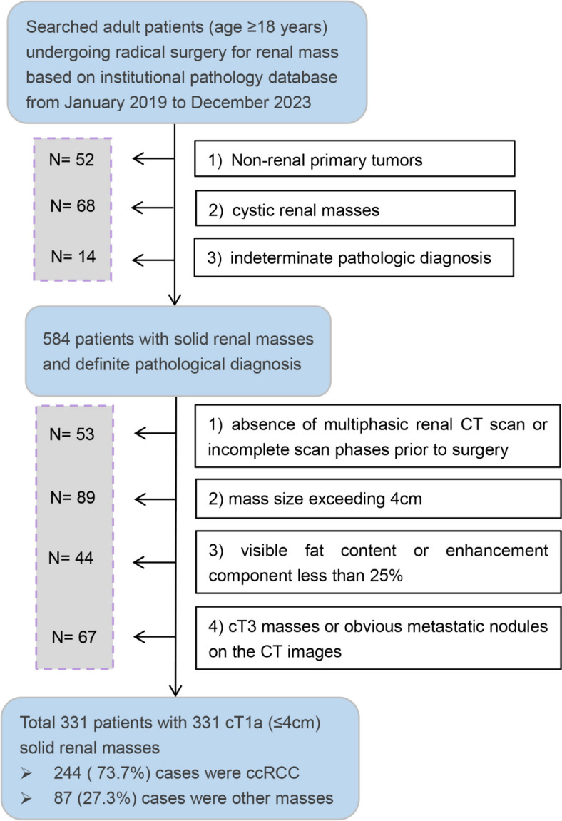
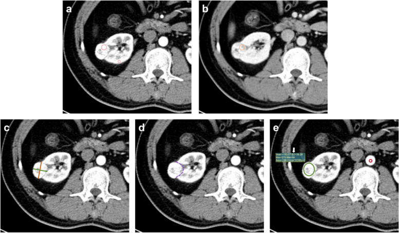
Methods: A retrospective collection of 331 patients with pathologically confirmed renal masses were enrolled in this study. Two radiologists independently assessed the CT images: in addition to heterogeneity score (HS) and mass-to-cortex corticomedullary attenuation ratio (MCAR), measured parameters included ratio of major diameter to minor diameter at the maximum axial section (Major axis / Minor axis), tumor-renal interface, standardized heterogeneity ratio (SHR), and standardized nephrographic reduction rate (SNRR). Spearman's correlation analysis was performed to evaluate the relationship between SHR and HS. Univariate and multivariate logistic regression analyses were employed to identify independent risk factors and then CT-score was adjusted by those indicators. The diagnostic efficacy of the modified CT-scores was evaluated using ROC curve analysis.
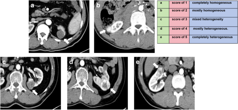
Results: The SHR and heterogeneity grade (HG) of mass were correlated positively with the HS (R = 0.749, 0.730, all P < 0.001). Logistic regression analysis determined that the Major axis / Minor axis (> 1.16), the tumor-renal interface (> 22.3 mm), and the SNRR (> 0.16) as additional independent risk factors to combine with HS and MCAR. Compared to the original CT-score, the two CT algorithms combined tumor-renal interface and SNRR showed significantly improved diagnostic efficacy for ccRCC (AUC: 0.770 vs. 0.861 and 0.862, all P < 0.001). The inter-observer agreement for HG was higher than that for HS (weighted Kappa coefficient: 0.797 vs. 0.722). The consistency of modified CT-score was also superior to original CT-score (weighted Kappa coefficient: 0.935 vs. 0.878).
Conclusion: The modified CT algorithms not only enhanced inter-reader consistency but also improved the diagnostic capability for ccRCC in small renal masses.
Cancer ImagingONCOLOGY-RADIOLOGY, NUCLEAR MEDICINE & MEDICAL IMAGING
CiteScore
7.00
自引率
0.00%
发文量
66
审稿时长
>12 weeks
期刊介绍:
Cancer Imaging is an open access, peer-reviewed journal publishing original articles, reviews and editorials written by expert international radiologists working in oncology.
The journal encompasses CT, MR, PET, ultrasound, radionuclide and multimodal imaging in all kinds of malignant tumours, plus new developments, techniques and innovations. Topics of interest include:
Breast Imaging
Chest
Complications of treatment
Ear, Nose & Throat
Gastrointestinal
Hepatobiliary & Pancreatic
Imaging biomarkers
Interventional
Lymphoma
Measurement of tumour response
Molecular functional imaging
Musculoskeletal
Neuro oncology
Nuclear Medicine
Paediatric.

 求助内容:
求助内容: 应助结果提醒方式:


