Viola Katharina Vetter, Perparim Limani, Falko Ensle, Michelle Leanne Brown, Lorenz Bankel, Marco Matteo Bühler, Chantal Pauli
{"title":"伴有广泛囊性改变的去分化脂肪肉瘤引起显著的诊断挑战:两例报告及文献复习。","authors":"Viola Katharina Vetter, Perparim Limani, Falko Ensle, Michelle Leanne Brown, Lorenz Bankel, Marco Matteo Bühler, Chantal Pauli","doi":"10.1186/s13000-025-01619-0","DOIUrl":null,"url":null,"abstract":"<p><strong>Background: </strong>Retroperitoneal dedifferentiated liposarcoma is a rare, aggressive malignancy, characterized by high rates of recurrences and the potential for metastasis. On imaging, these tumors typically present as a solid mass with lipomatous and non-lipomatous components. Cystic changes of dedifferentiated liposarcomas is exceedingly rare and might pose significant diagnostic challenges, with only a few cases reported in the literature. REPORT OF 2 CASES: We here present two cases of retroperitoneal dedifferentiated liposarcoma with a rare cystic presentation in two female patients aged 51 and 62 years. Imaging revealed large perinephric cystic masses measuring up to 13.0 cm and 16.1 cm, respectively, with calcifications of the cyst wall observed in the second case. Differential diagnoses included cystic echinococcosis, mesenchymal neoplasms, and benign cystic lesions (e.g. endometrial cyst). Both patients underwent upfront compartmental en-bloc surgical resection of the tumor mass and the kidney after multidisciplinary tumor board (MDT) discussion. Macroscopically, the tumors were adherent to but sharply demarcated from the kidney. Histological examination of the first case revealed a small component of well-differentiated liposarcoma (WDLPS) adjacent to a large non-lipogenic sarcoma with a prominent whirling pattern, compatible with dedifferentiation. The second case demonstrated a spindle cell neoplasm with prominent osteosarcomatous heterologous differentiation. MDM2 amplification was confirmed in both cases by molecular testing. No long-term follow-up data is available for either patient.</p><p><strong>Conclusion: </strong>In conclusion, these cases highlight the importance of recognizing unusual and extensive cystic changes of dedifferentiated liposarcoma, which can complicate the diagnostic work-up.</p>","PeriodicalId":11237,"journal":{"name":"Diagnostic Pathology","volume":"20 1","pages":"23"},"PeriodicalIF":2.3000,"publicationDate":"2025-02-27","publicationTypes":"Journal Article","fieldsOfStudy":null,"isOpenAccess":false,"openAccessPdf":"https://www.ncbi.nlm.nih.gov/pmc/articles/PMC11866618/pdf/","citationCount":"0","resultStr":"{\"title\":\"Dedifferentiated liposarcoma with extensive cystic change causing significant diagnostic challenges: report of two cases and review of the literature.\",\"authors\":\"Viola Katharina Vetter, Perparim Limani, Falko Ensle, Michelle Leanne Brown, Lorenz Bankel, Marco Matteo Bühler, Chantal Pauli\",\"doi\":\"10.1186/s13000-025-01619-0\",\"DOIUrl\":null,\"url\":null,\"abstract\":\"<p><strong>Background: </strong>Retroperitoneal dedifferentiated liposarcoma is a rare, aggressive malignancy, characterized by high rates of recurrences and the potential for metastasis. On imaging, these tumors typically present as a solid mass with lipomatous and non-lipomatous components. Cystic changes of dedifferentiated liposarcomas is exceedingly rare and might pose significant diagnostic challenges, with only a few cases reported in the literature. REPORT OF 2 CASES: We here present two cases of retroperitoneal dedifferentiated liposarcoma with a rare cystic presentation in two female patients aged 51 and 62 years. Imaging revealed large perinephric cystic masses measuring up to 13.0 cm and 16.1 cm, respectively, with calcifications of the cyst wall observed in the second case. Differential diagnoses included cystic echinococcosis, mesenchymal neoplasms, and benign cystic lesions (e.g. endometrial cyst). Both patients underwent upfront compartmental en-bloc surgical resection of the tumor mass and the kidney after multidisciplinary tumor board (MDT) discussion. Macroscopically, the tumors were adherent to but sharply demarcated from the kidney. Histological examination of the first case revealed a small component of well-differentiated liposarcoma (WDLPS) adjacent to a large non-lipogenic sarcoma with a prominent whirling pattern, compatible with dedifferentiation. The second case demonstrated a spindle cell neoplasm with prominent osteosarcomatous heterologous differentiation. MDM2 amplification was confirmed in both cases by molecular testing. No long-term follow-up data is available for either patient.</p><p><strong>Conclusion: </strong>In conclusion, these cases highlight the importance of recognizing unusual and extensive cystic changes of dedifferentiated liposarcoma, which can complicate the diagnostic work-up.</p>\",\"PeriodicalId\":11237,\"journal\":{\"name\":\"Diagnostic Pathology\",\"volume\":\"20 1\",\"pages\":\"23\"},\"PeriodicalIF\":2.3000,\"publicationDate\":\"2025-02-27\",\"publicationTypes\":\"Journal Article\",\"fieldsOfStudy\":null,\"isOpenAccess\":false,\"openAccessPdf\":\"https://www.ncbi.nlm.nih.gov/pmc/articles/PMC11866618/pdf/\",\"citationCount\":\"0\",\"resultStr\":null,\"platform\":\"Semanticscholar\",\"paperid\":null,\"PeriodicalName\":\"Diagnostic Pathology\",\"FirstCategoryId\":\"3\",\"ListUrlMain\":\"https://doi.org/10.1186/s13000-025-01619-0\",\"RegionNum\":3,\"RegionCategory\":\"医学\",\"ArticlePicture\":[],\"TitleCN\":null,\"AbstractTextCN\":null,\"PMCID\":null,\"EPubDate\":\"\",\"PubModel\":\"\",\"JCR\":\"Q2\",\"JCRName\":\"PATHOLOGY\",\"Score\":null,\"Total\":0}","platform":"Semanticscholar","paperid":null,"PeriodicalName":"Diagnostic Pathology","FirstCategoryId":"3","ListUrlMain":"https://doi.org/10.1186/s13000-025-01619-0","RegionNum":3,"RegionCategory":"医学","ArticlePicture":[],"TitleCN":null,"AbstractTextCN":null,"PMCID":null,"EPubDate":"","PubModel":"","JCR":"Q2","JCRName":"PATHOLOGY","Score":null,"Total":0}
Dedifferentiated liposarcoma with extensive cystic change causing significant diagnostic challenges: report of two cases and review of the literature.
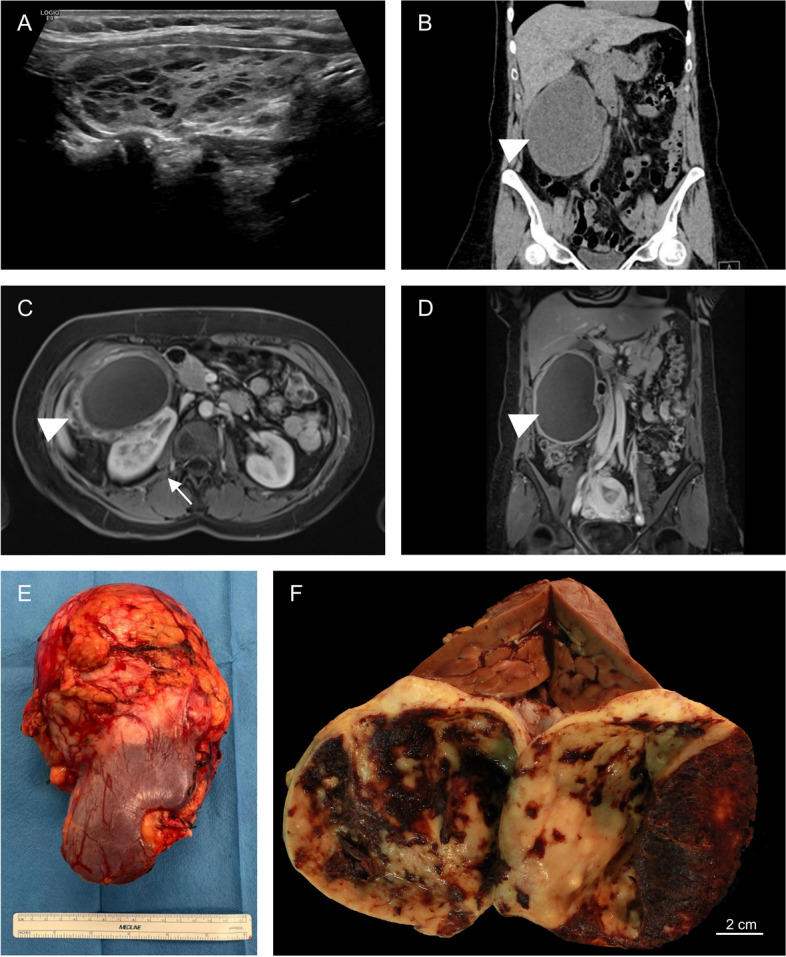
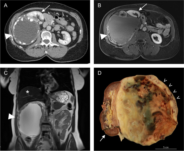
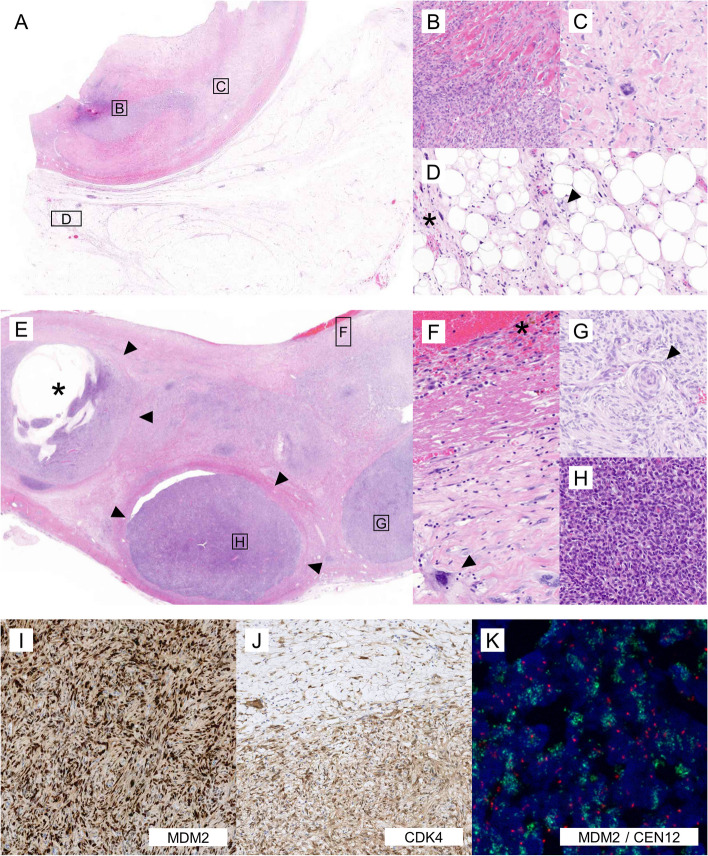
Background: Retroperitoneal dedifferentiated liposarcoma is a rare, aggressive malignancy, characterized by high rates of recurrences and the potential for metastasis. On imaging, these tumors typically present as a solid mass with lipomatous and non-lipomatous components. Cystic changes of dedifferentiated liposarcomas is exceedingly rare and might pose significant diagnostic challenges, with only a few cases reported in the literature. REPORT OF 2 CASES: We here present two cases of retroperitoneal dedifferentiated liposarcoma with a rare cystic presentation in two female patients aged 51 and 62 years. Imaging revealed large perinephric cystic masses measuring up to 13.0 cm and 16.1 cm, respectively, with calcifications of the cyst wall observed in the second case. Differential diagnoses included cystic echinococcosis, mesenchymal neoplasms, and benign cystic lesions (e.g. endometrial cyst). Both patients underwent upfront compartmental en-bloc surgical resection of the tumor mass and the kidney after multidisciplinary tumor board (MDT) discussion. Macroscopically, the tumors were adherent to but sharply demarcated from the kidney. Histological examination of the first case revealed a small component of well-differentiated liposarcoma (WDLPS) adjacent to a large non-lipogenic sarcoma with a prominent whirling pattern, compatible with dedifferentiation. The second case demonstrated a spindle cell neoplasm with prominent osteosarcomatous heterologous differentiation. MDM2 amplification was confirmed in both cases by molecular testing. No long-term follow-up data is available for either patient.
Conclusion: In conclusion, these cases highlight the importance of recognizing unusual and extensive cystic changes of dedifferentiated liposarcoma, which can complicate the diagnostic work-up.
期刊介绍:
Diagnostic Pathology is an open access, peer-reviewed, online journal that considers research in surgical and clinical pathology, immunology, and biology, with a special focus on cutting-edge approaches in diagnostic pathology and tissue-based therapy. The journal covers all aspects of surgical pathology, including classic diagnostic pathology, prognosis-related diagnosis (tumor stages, prognosis markers, such as MIB-percentage, hormone receptors, etc.), and therapy-related findings. The journal also focuses on the technological aspects of pathology, including molecular biology techniques, morphometry aspects (stereology, DNA analysis, syntactic structure analysis), communication aspects (telecommunication, virtual microscopy, virtual pathology institutions, etc.), and electronic education and quality assurance (for example interactive publication, on-line references with automated updating, etc.).

 求助内容:
求助内容: 应助结果提醒方式:


