{"title":"食管癌伴右上肺静脉异常胸腔镜食管切除术伴隆突下淋巴结清扫术中吲哚菁绿荧光显像1例并文献复习。","authors":"Naoto Ujiie, Takanobu Nakamura, Takahiro Heishi, Yusuke Taniyama, Takashi Kamei","doi":"10.5761/atcs.cr.25-00015","DOIUrl":null,"url":null,"abstract":"<p><p>A 68-year-old woman was diagnosed with clinical T3N1M0 middle thoracic esophageal cancer. Preoperative three-dimensional computed tomography indicated a right superior posterior pulmonary vein (RSPPV) anomaly, which ran behind the right intermediate bronchus. The patient underwent thoracoscopic esophagectomy with mediastinal lymph node (LN) dissection. Before we began the dissection of the right subcarinal LN, we administered indocyanine green intravenously to confirm the running position of the anomalous RSPPV, and we were able to ascertain its placement accurately with correct recognition of the difference between the blood vessels and surrounding tissue. Although the patient had LN metastasis adjacent to this anomalous vessel and the dissection procedure was tough due to tightly adhesion, intraoperative fluorescent imaging enabled us to perform the dissection without any superfluous vascular injury. Intraoperative fluorescent imaging is very useful in such cases, providing accurate intraoperative information on the location of the anomaly and facilitating safer surgery.</p>","PeriodicalId":93877,"journal":{"name":"Annals of thoracic and cardiovascular surgery : official journal of the Association of Thoracic and Cardiovascular Surgeons of Asia","volume":"31 1","pages":""},"PeriodicalIF":1.3000,"publicationDate":"2025-01-01","publicationTypes":"Journal Article","fieldsOfStudy":null,"isOpenAccess":false,"openAccessPdf":"https://www.ncbi.nlm.nih.gov/pmc/articles/PMC11891391/pdf/","citationCount":"0","resultStr":"{\"title\":\"Intraoperative Fluorescent Imaging with Indocyanine Green during Thoracoscopic Esophagectomy with Subcarinal Lymph Node Dissection for Esophageal Cancer with a Right Superior Pulmonary Vein Anomaly: A Case Report and Literature Review.\",\"authors\":\"Naoto Ujiie, Takanobu Nakamura, Takahiro Heishi, Yusuke Taniyama, Takashi Kamei\",\"doi\":\"10.5761/atcs.cr.25-00015\",\"DOIUrl\":null,\"url\":null,\"abstract\":\"<p><p>A 68-year-old woman was diagnosed with clinical T3N1M0 middle thoracic esophageal cancer. Preoperative three-dimensional computed tomography indicated a right superior posterior pulmonary vein (RSPPV) anomaly, which ran behind the right intermediate bronchus. The patient underwent thoracoscopic esophagectomy with mediastinal lymph node (LN) dissection. Before we began the dissection of the right subcarinal LN, we administered indocyanine green intravenously to confirm the running position of the anomalous RSPPV, and we were able to ascertain its placement accurately with correct recognition of the difference between the blood vessels and surrounding tissue. Although the patient had LN metastasis adjacent to this anomalous vessel and the dissection procedure was tough due to tightly adhesion, intraoperative fluorescent imaging enabled us to perform the dissection without any superfluous vascular injury. Intraoperative fluorescent imaging is very useful in such cases, providing accurate intraoperative information on the location of the anomaly and facilitating safer surgery.</p>\",\"PeriodicalId\":93877,\"journal\":{\"name\":\"Annals of thoracic and cardiovascular surgery : official journal of the Association of Thoracic and Cardiovascular Surgeons of Asia\",\"volume\":\"31 1\",\"pages\":\"\"},\"PeriodicalIF\":1.3000,\"publicationDate\":\"2025-01-01\",\"publicationTypes\":\"Journal Article\",\"fieldsOfStudy\":null,\"isOpenAccess\":false,\"openAccessPdf\":\"https://www.ncbi.nlm.nih.gov/pmc/articles/PMC11891391/pdf/\",\"citationCount\":\"0\",\"resultStr\":null,\"platform\":\"Semanticscholar\",\"paperid\":null,\"PeriodicalName\":\"Annals of thoracic and cardiovascular surgery : official journal of the Association of Thoracic and Cardiovascular Surgeons of Asia\",\"FirstCategoryId\":\"1085\",\"ListUrlMain\":\"https://doi.org/10.5761/atcs.cr.25-00015\",\"RegionNum\":0,\"RegionCategory\":null,\"ArticlePicture\":[],\"TitleCN\":null,\"AbstractTextCN\":null,\"PMCID\":null,\"EPubDate\":\"\",\"PubModel\":\"\",\"JCR\":\"\",\"JCRName\":\"\",\"Score\":null,\"Total\":0}","platform":"Semanticscholar","paperid":null,"PeriodicalName":"Annals of thoracic and cardiovascular surgery : official journal of the Association of Thoracic and Cardiovascular Surgeons of Asia","FirstCategoryId":"1085","ListUrlMain":"https://doi.org/10.5761/atcs.cr.25-00015","RegionNum":0,"RegionCategory":null,"ArticlePicture":[],"TitleCN":null,"AbstractTextCN":null,"PMCID":null,"EPubDate":"","PubModel":"","JCR":"","JCRName":"","Score":null,"Total":0}
Intraoperative Fluorescent Imaging with Indocyanine Green during Thoracoscopic Esophagectomy with Subcarinal Lymph Node Dissection for Esophageal Cancer with a Right Superior Pulmonary Vein Anomaly: A Case Report and Literature Review.
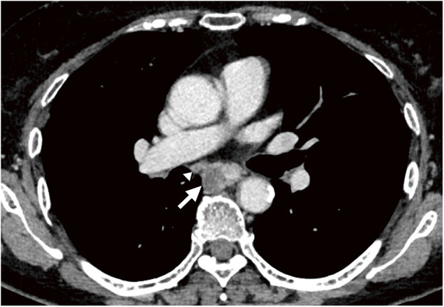
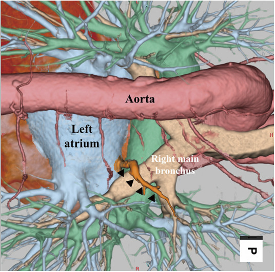
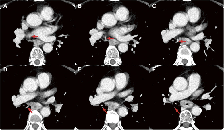
A 68-year-old woman was diagnosed with clinical T3N1M0 middle thoracic esophageal cancer. Preoperative three-dimensional computed tomography indicated a right superior posterior pulmonary vein (RSPPV) anomaly, which ran behind the right intermediate bronchus. The patient underwent thoracoscopic esophagectomy with mediastinal lymph node (LN) dissection. Before we began the dissection of the right subcarinal LN, we administered indocyanine green intravenously to confirm the running position of the anomalous RSPPV, and we were able to ascertain its placement accurately with correct recognition of the difference between the blood vessels and surrounding tissue. Although the patient had LN metastasis adjacent to this anomalous vessel and the dissection procedure was tough due to tightly adhesion, intraoperative fluorescent imaging enabled us to perform the dissection without any superfluous vascular injury. Intraoperative fluorescent imaging is very useful in such cases, providing accurate intraoperative information on the location of the anomaly and facilitating safer surgery.

 求助内容:
求助内容: 应助结果提醒方式:


