Laura DiChiacchio, Ikeoluwapo Ibrahim, Alison Grazioli, Neda Bionghi, Kiran Batra, Sameer Chabbra, Nicholas Ladikos, Vaidehi Kaza, Srinivas Bollineni, Manish R Mohanka, Adrian Lawrence, Fernando Torres, Aldo Iacono, Daniel Herr, Irina Timofte
{"title":"随着时间的推移,放射学表现的改善与需要体外膜氧合的病毒性ARDS患者的生存率增加有关。","authors":"Laura DiChiacchio, Ikeoluwapo Ibrahim, Alison Grazioli, Neda Bionghi, Kiran Batra, Sameer Chabbra, Nicholas Ladikos, Vaidehi Kaza, Srinivas Bollineni, Manish R Mohanka, Adrian Lawrence, Fernando Torres, Aldo Iacono, Daniel Herr, Irina Timofte","doi":"10.4103/lungindia.lungindia_501_24","DOIUrl":null,"url":null,"abstract":"<p><strong>Introduction: </strong>Acute respiratory distress syndrome (ARDS) requiring venovenous extracorporeal membrane oxygenation (VV ECMO) support is associated with chest radiograph changes commonly referred to as \"drowning ECMO lung\" ECMO lung presents as white-out of both lung fields, involving all lobes of the bilateral lungs. While the clinical significance of chest radiograph findings over time has been described in the general ARDS population, it has not been evaluated specifically in VV ECMO patients. This subpopulation suffers the most severe disease as well as the confounding effects of ECMO support.</p><p><strong>Materials and methods: </strong>We identified 28 patients requiring VV ECMO cannulation for influenza-related ARDS between September 2009 and January 2018. Interpretation of chest X-ray images was divided into zones that correspond to anatomical lobes on computed tomography. Progression of radiologic injury was assessed by analysing the number of zones involved on the chest radiograph (X-ray) at days 1, 3, 7, 14, and 21 from cannulation and discharge. The primary endpoint was survival to hospital discharge.</p><p><strong>Results: </strong>The majority of patients had complete opacification on days 1, 3, and 7 after VV ECMO cannulation. Patients with persistent complete opacification on chest X-ray infiltrate by day 14, following cannulation had an increased mortality. Survival to hospital discharge was increased in patients demonstrating improvement in radiological findings at day 19 compared to patients without significant radiologic improvement (100% vs 53%, log-rank P = 0.003).</p><p><strong>Conclusion: </strong>The evolution and recovery of lung injury reflected by serial chest X-ray imaging studies after influenza-related ARDS requiring VV ECMO support is associated with improved survival in this single centre, retrospective cohort.</p>","PeriodicalId":47462,"journal":{"name":"Lung India","volume":"42 2","pages":"91-96"},"PeriodicalIF":1.2000,"publicationDate":"2025-03-01","publicationTypes":"Journal Article","fieldsOfStudy":null,"isOpenAccess":false,"openAccessPdf":"https://www.ncbi.nlm.nih.gov/pmc/articles/PMC11952723/pdf/","citationCount":"0","resultStr":"{\"title\":\"Improvement in radiologic findings over time is associated with increased survival in patients with viral ARDS requiring extracorporeal membrane oxygenation.\",\"authors\":\"Laura DiChiacchio, Ikeoluwapo Ibrahim, Alison Grazioli, Neda Bionghi, Kiran Batra, Sameer Chabbra, Nicholas Ladikos, Vaidehi Kaza, Srinivas Bollineni, Manish R Mohanka, Adrian Lawrence, Fernando Torres, Aldo Iacono, Daniel Herr, Irina Timofte\",\"doi\":\"10.4103/lungindia.lungindia_501_24\",\"DOIUrl\":null,\"url\":null,\"abstract\":\"<p><strong>Introduction: </strong>Acute respiratory distress syndrome (ARDS) requiring venovenous extracorporeal membrane oxygenation (VV ECMO) support is associated with chest radiograph changes commonly referred to as \\\"drowning ECMO lung\\\" ECMO lung presents as white-out of both lung fields, involving all lobes of the bilateral lungs. While the clinical significance of chest radiograph findings over time has been described in the general ARDS population, it has not been evaluated specifically in VV ECMO patients. This subpopulation suffers the most severe disease as well as the confounding effects of ECMO support.</p><p><strong>Materials and methods: </strong>We identified 28 patients requiring VV ECMO cannulation for influenza-related ARDS between September 2009 and January 2018. Interpretation of chest X-ray images was divided into zones that correspond to anatomical lobes on computed tomography. Progression of radiologic injury was assessed by analysing the number of zones involved on the chest radiograph (X-ray) at days 1, 3, 7, 14, and 21 from cannulation and discharge. The primary endpoint was survival to hospital discharge.</p><p><strong>Results: </strong>The majority of patients had complete opacification on days 1, 3, and 7 after VV ECMO cannulation. Patients with persistent complete opacification on chest X-ray infiltrate by day 14, following cannulation had an increased mortality. Survival to hospital discharge was increased in patients demonstrating improvement in radiological findings at day 19 compared to patients without significant radiologic improvement (100% vs 53%, log-rank P = 0.003).</p><p><strong>Conclusion: </strong>The evolution and recovery of lung injury reflected by serial chest X-ray imaging studies after influenza-related ARDS requiring VV ECMO support is associated with improved survival in this single centre, retrospective cohort.</p>\",\"PeriodicalId\":47462,\"journal\":{\"name\":\"Lung India\",\"volume\":\"42 2\",\"pages\":\"91-96\"},\"PeriodicalIF\":1.2000,\"publicationDate\":\"2025-03-01\",\"publicationTypes\":\"Journal Article\",\"fieldsOfStudy\":null,\"isOpenAccess\":false,\"openAccessPdf\":\"https://www.ncbi.nlm.nih.gov/pmc/articles/PMC11952723/pdf/\",\"citationCount\":\"0\",\"resultStr\":null,\"platform\":\"Semanticscholar\",\"paperid\":null,\"PeriodicalName\":\"Lung India\",\"FirstCategoryId\":\"1085\",\"ListUrlMain\":\"https://doi.org/10.4103/lungindia.lungindia_501_24\",\"RegionNum\":0,\"RegionCategory\":null,\"ArticlePicture\":[],\"TitleCN\":null,\"AbstractTextCN\":null,\"PMCID\":null,\"EPubDate\":\"2025/2/27 0:00:00\",\"PubModel\":\"Epub\",\"JCR\":\"Q4\",\"JCRName\":\"RESPIRATORY SYSTEM\",\"Score\":null,\"Total\":0}","platform":"Semanticscholar","paperid":null,"PeriodicalName":"Lung India","FirstCategoryId":"1085","ListUrlMain":"https://doi.org/10.4103/lungindia.lungindia_501_24","RegionNum":0,"RegionCategory":null,"ArticlePicture":[],"TitleCN":null,"AbstractTextCN":null,"PMCID":null,"EPubDate":"2025/2/27 0:00:00","PubModel":"Epub","JCR":"Q4","JCRName":"RESPIRATORY SYSTEM","Score":null,"Total":0}
引用次数: 0
摘要
简介:需要静脉静脉体外膜氧合(VV ECMO)支持的急性呼吸窘迫综合征(ARDS)与胸片改变有关,通常被称为“溺水ECMO肺”。ECMO肺表现为双肺野白化,累及双肺的所有肺叶。虽然在一般ARDS人群中胸片表现随时间变化的临床意义已被描述,但尚未对VV ECMO患者进行专门评估。这个亚群遭受最严重的疾病以及ECMO支持的混淆效应。材料和方法:我们确定了2009年9月至2018年1月期间需要VV ECMO插管治疗流感相关ARDS的28例患者。胸部x线图像的解释被划分为与计算机断层扫描上的解剖叶相对应的区域。通过分析插管和出院后第1、3、7、14和21天胸片(x线)上受累区域的数量来评估放射学损伤的进展。主要终点是生存至出院。结果:大多数患者在VV ECMO插管后第1,3,7天出现完全混浊。插管后第14天胸片浸润持续完全混浊的患者死亡率增加。与放射学无显著改善的患者相比,在第19天放射学表现改善的患者到出院的生存率增加(100% vs 53%, log-rank P = 0.003)。结论:在单中心、回顾性队列研究中,需要VV ECMO支持的流感相关ARDS患者的肺部损伤的演变和恢复与生存率的提高有关。
Improvement in radiologic findings over time is associated with increased survival in patients with viral ARDS requiring extracorporeal membrane oxygenation.
Introduction: Acute respiratory distress syndrome (ARDS) requiring venovenous extracorporeal membrane oxygenation (VV ECMO) support is associated with chest radiograph changes commonly referred to as "drowning ECMO lung" ECMO lung presents as white-out of both lung fields, involving all lobes of the bilateral lungs. While the clinical significance of chest radiograph findings over time has been described in the general ARDS population, it has not been evaluated specifically in VV ECMO patients. This subpopulation suffers the most severe disease as well as the confounding effects of ECMO support.
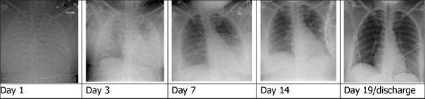
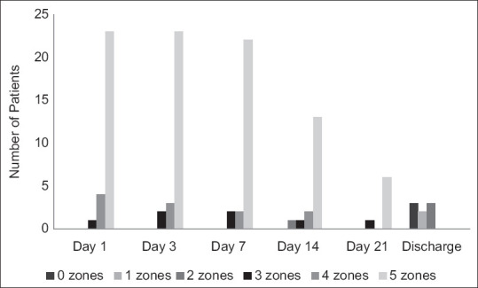
Materials and methods: We identified 28 patients requiring VV ECMO cannulation for influenza-related ARDS between September 2009 and January 2018. Interpretation of chest X-ray images was divided into zones that correspond to anatomical lobes on computed tomography. Progression of radiologic injury was assessed by analysing the number of zones involved on the chest radiograph (X-ray) at days 1, 3, 7, 14, and 21 from cannulation and discharge. The primary endpoint was survival to hospital discharge.
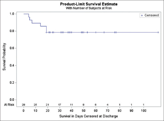
Results: The majority of patients had complete opacification on days 1, 3, and 7 after VV ECMO cannulation. Patients with persistent complete opacification on chest X-ray infiltrate by day 14, following cannulation had an increased mortality. Survival to hospital discharge was increased in patients demonstrating improvement in radiological findings at day 19 compared to patients without significant radiologic improvement (100% vs 53%, log-rank P = 0.003).
Conclusion: The evolution and recovery of lung injury reflected by serial chest X-ray imaging studies after influenza-related ARDS requiring VV ECMO support is associated with improved survival in this single centre, retrospective cohort.

 求助内容:
求助内容: 应助结果提醒方式:


