{"title":"血管内皮生长因子(VEGF)水平在伴有腹水的胃肠道肿瘤中的诊断价值-横断面研究。","authors":"Evangeline Mary Kiruba Samuel, Sudharsanan Sundaramurthi, Nandeesha Hanumanthappa, Vishnu Prasad Nelamangalaramakrishnaiah","doi":"10.47717/turkjsurg.2025.6592","DOIUrl":null,"url":null,"abstract":"<p><strong>Objective: </strong>Malignant ascites is suggestive of peritoneal carcinomatosis. The distinction between malignant and non-malignant ascites in a patient with malignancy is important, as it alters the management and prognosis. Current diagnostic methods are imaging, cytology, and diagnostic laparoscopy, all of which have low sensitivities. The vascular endothelial growth factor (VEGF) is essential for tumour growth and, hence, ascitic VEGF levels can be a diagnostic method for malignant ascites.</p><p><strong>Material and methods: </strong>This cross-sectional study was conducted in patients with gastrointestinal malignancies and ascites. The calculated sample size was 68 patients, who were divided into those who were truly positive or negative for malignant ascites based on a composite gold standard, comprising cytology, contrast enhanced computed tomography, and laparoscopy. The ascitic VEGF levels in these patients were compared.</p><p><strong>Results: </strong>A total of 84 patients were enrolled, of whom 60.71% were found to have malignant ascites. It was found that the greater the volume of ascites, the greater the statistical likelihood of finding truly malignant ascites. The ascitic VEGF levels had a non-normal distribution, with median values of 783.64 and 41.12 pg/mL for malignant and non-malignant ascites (p<0.001). Using a receiver operating characteristics curve, a cut-off of 83.68 pg/mL was obtained, with a sensitivity of 100% and a specificity of 93.94%.</p><p><strong>Conclusion: </strong>This study demonstrates that ascitic VEGF levels are significantly elevated in patients with gastrointestinal malignancies and malignant ascites and hence can reliably be used for diagnosing malignant ascites. This study also shows that massive ascites and well-differentiated tumours have a higher rate of peritoneal carcinomatosis.</p>","PeriodicalId":23374,"journal":{"name":"Turkish Journal of Surgery","volume":"41 1","pages":"78-84"},"PeriodicalIF":0.6000,"publicationDate":"2025-02-27","publicationTypes":"Journal Article","fieldsOfStudy":null,"isOpenAccess":false,"openAccessPdf":"https://www.ncbi.nlm.nih.gov/pmc/articles/PMC11878185/pdf/","citationCount":"0","resultStr":"{\"title\":\"Diagnostic value of vascular endothelial growth factor (VEGF) levels in gastrointestinal cancers with ascites - A cross sectional study.\",\"authors\":\"Evangeline Mary Kiruba Samuel, Sudharsanan Sundaramurthi, Nandeesha Hanumanthappa, Vishnu Prasad Nelamangalaramakrishnaiah\",\"doi\":\"10.47717/turkjsurg.2025.6592\",\"DOIUrl\":null,\"url\":null,\"abstract\":\"<p><strong>Objective: </strong>Malignant ascites is suggestive of peritoneal carcinomatosis. The distinction between malignant and non-malignant ascites in a patient with malignancy is important, as it alters the management and prognosis. Current diagnostic methods are imaging, cytology, and diagnostic laparoscopy, all of which have low sensitivities. The vascular endothelial growth factor (VEGF) is essential for tumour growth and, hence, ascitic VEGF levels can be a diagnostic method for malignant ascites.</p><p><strong>Material and methods: </strong>This cross-sectional study was conducted in patients with gastrointestinal malignancies and ascites. The calculated sample size was 68 patients, who were divided into those who were truly positive or negative for malignant ascites based on a composite gold standard, comprising cytology, contrast enhanced computed tomography, and laparoscopy. The ascitic VEGF levels in these patients were compared.</p><p><strong>Results: </strong>A total of 84 patients were enrolled, of whom 60.71% were found to have malignant ascites. It was found that the greater the volume of ascites, the greater the statistical likelihood of finding truly malignant ascites. The ascitic VEGF levels had a non-normal distribution, with median values of 783.64 and 41.12 pg/mL for malignant and non-malignant ascites (p<0.001). Using a receiver operating characteristics curve, a cut-off of 83.68 pg/mL was obtained, with a sensitivity of 100% and a specificity of 93.94%.</p><p><strong>Conclusion: </strong>This study demonstrates that ascitic VEGF levels are significantly elevated in patients with gastrointestinal malignancies and malignant ascites and hence can reliably be used for diagnosing malignant ascites. This study also shows that massive ascites and well-differentiated tumours have a higher rate of peritoneal carcinomatosis.</p>\",\"PeriodicalId\":23374,\"journal\":{\"name\":\"Turkish Journal of Surgery\",\"volume\":\"41 1\",\"pages\":\"78-84\"},\"PeriodicalIF\":0.6000,\"publicationDate\":\"2025-02-27\",\"publicationTypes\":\"Journal Article\",\"fieldsOfStudy\":null,\"isOpenAccess\":false,\"openAccessPdf\":\"https://www.ncbi.nlm.nih.gov/pmc/articles/PMC11878185/pdf/\",\"citationCount\":\"0\",\"resultStr\":null,\"platform\":\"Semanticscholar\",\"paperid\":null,\"PeriodicalName\":\"Turkish Journal of Surgery\",\"FirstCategoryId\":\"1085\",\"ListUrlMain\":\"https://doi.org/10.47717/turkjsurg.2025.6592\",\"RegionNum\":0,\"RegionCategory\":null,\"ArticlePicture\":[],\"TitleCN\":null,\"AbstractTextCN\":null,\"PMCID\":null,\"EPubDate\":\"\",\"PubModel\":\"\",\"JCR\":\"Q4\",\"JCRName\":\"SURGERY\",\"Score\":null,\"Total\":0}","platform":"Semanticscholar","paperid":null,"PeriodicalName":"Turkish Journal of Surgery","FirstCategoryId":"1085","ListUrlMain":"https://doi.org/10.47717/turkjsurg.2025.6592","RegionNum":0,"RegionCategory":null,"ArticlePicture":[],"TitleCN":null,"AbstractTextCN":null,"PMCID":null,"EPubDate":"","PubModel":"","JCR":"Q4","JCRName":"SURGERY","Score":null,"Total":0}
Diagnostic value of vascular endothelial growth factor (VEGF) levels in gastrointestinal cancers with ascites - A cross sectional study.
Objective: Malignant ascites is suggestive of peritoneal carcinomatosis. The distinction between malignant and non-malignant ascites in a patient with malignancy is important, as it alters the management and prognosis. Current diagnostic methods are imaging, cytology, and diagnostic laparoscopy, all of which have low sensitivities. The vascular endothelial growth factor (VEGF) is essential for tumour growth and, hence, ascitic VEGF levels can be a diagnostic method for malignant ascites.
Material and methods: This cross-sectional study was conducted in patients with gastrointestinal malignancies and ascites. The calculated sample size was 68 patients, who were divided into those who were truly positive or negative for malignant ascites based on a composite gold standard, comprising cytology, contrast enhanced computed tomography, and laparoscopy. The ascitic VEGF levels in these patients were compared.
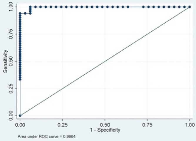
Results: A total of 84 patients were enrolled, of whom 60.71% were found to have malignant ascites. It was found that the greater the volume of ascites, the greater the statistical likelihood of finding truly malignant ascites. The ascitic VEGF levels had a non-normal distribution, with median values of 783.64 and 41.12 pg/mL for malignant and non-malignant ascites (p<0.001). Using a receiver operating characteristics curve, a cut-off of 83.68 pg/mL was obtained, with a sensitivity of 100% and a specificity of 93.94%.
Conclusion: This study demonstrates that ascitic VEGF levels are significantly elevated in patients with gastrointestinal malignancies and malignant ascites and hence can reliably be used for diagnosing malignant ascites. This study also shows that massive ascites and well-differentiated tumours have a higher rate of peritoneal carcinomatosis.

 求助内容:
求助内容: 应助结果提醒方式:


