磁共振引导下自监督PET重建的临床与深度学习评价。

IF 3.5 Q1 RADIOLOGY, NUCLEAR MEDICINE & MEDICAL IMAGING
Jessica B. Hopson;Sam Ellis;Anthime Flaus;Colm J. McGinnity;Radhouene Neji;Andrew J. Reader;Alexander Hammers
{"title":"磁共振引导下自监督PET重建的临床与深度学习评价。","authors":"Jessica B. Hopson;Sam Ellis;Anthime Flaus;Colm J. McGinnity;Radhouene Neji;Andrew J. Reader;Alexander Hammers","doi":"10.1109/TRPMS.2024.3496779","DOIUrl":null,"url":null,"abstract":"Reduced dose positron emission tomography (PET) lowers the radiation dose to patients and reduces costs. Lower-count data, however, degrades reconstructed image quality. Advanced reconstruction methods help mitigate image quality losses, but it is important to assess the resulting images from a clinical perspective. Two experienced clinicians assessed four PET reconstruction algorithms for [18F]FDG brain data, compared to a clinical standard reference (maximum-likelihood expectation-maximization (MLEM)), based on seven clinical image quality metrics: global quality rating, pattern recognition, diagnostic confidence (all on a scale of 0–4), sharpness, caudate-putamen separation (CP), noise, and contrast (on a scale between 0–2). The reconstruction methods assessed were a guided and unguided version of self-supervised maximum a posteriori EM (MAPEM) (where the guidance case used the patient’s MR image to control the smoothness penalty). For 3 of the 11 patient datasets reconstructed, post-smoothed versions of the MAPEM reconstruction were also considered, where the smoothing was with the point-spread-function used in the resolution modelling. Statistically significant improvements were observed in sharpness, CP, and contrast for self-supervised MR-guided MAPEM compared to MLEM. For example, MLEM scored between 1-1.1 out of 2 for sharpness, CP, and contrast, whereas self-supervised MR-guided MAPEM scored between 1.5-1.75. In addition to the clinical evaluation, pretrained convolutional neural networks (CNNs) were used to assess the image quality of a further 62 images. The CNNs demonstrated similar trends to the clinician, showing their potential as automated standalone observers. Both the clinical and CNN assessments suggest when using only 5% of the standard injected dose, self-supervised MR-guided MAPEM reconstruction matches the 100% MLEM case for overall performance. This makes the images far more clinically useful than standard MLEM.","PeriodicalId":46807,"journal":{"name":"IEEE Transactions on Radiation and Plasma Medical Sciences","volume":"9 3","pages":"337-346"},"PeriodicalIF":3.5000,"publicationDate":"2024-11-15","publicationTypes":"Journal Article","fieldsOfStudy":null,"isOpenAccess":false,"openAccessPdf":"","citationCount":"0","resultStr":"{\"title\":\"Clinical and Deep-Learned Evaluation of MR-Guided Self-Supervised PET Reconstruction\",\"authors\":\"Jessica B. Hopson;Sam Ellis;Anthime Flaus;Colm J. McGinnity;Radhouene Neji;Andrew J. Reader;Alexander Hammers\",\"doi\":\"10.1109/TRPMS.2024.3496779\",\"DOIUrl\":null,\"url\":null,\"abstract\":\"Reduced dose positron emission tomography (PET) lowers the radiation dose to patients and reduces costs. Lower-count data, however, degrades reconstructed image quality. Advanced reconstruction methods help mitigate image quality losses, but it is important to assess the resulting images from a clinical perspective. Two experienced clinicians assessed four PET reconstruction algorithms for [18F]FDG brain data, compared to a clinical standard reference (maximum-likelihood expectation-maximization (MLEM)), based on seven clinical image quality metrics: global quality rating, pattern recognition, diagnostic confidence (all on a scale of 0–4), sharpness, caudate-putamen separation (CP), noise, and contrast (on a scale between 0–2). The reconstruction methods assessed were a guided and unguided version of self-supervised maximum a posteriori EM (MAPEM) (where the guidance case used the patient’s MR image to control the smoothness penalty). For 3 of the 11 patient datasets reconstructed, post-smoothed versions of the MAPEM reconstruction were also considered, where the smoothing was with the point-spread-function used in the resolution modelling. Statistically significant improvements were observed in sharpness, CP, and contrast for self-supervised MR-guided MAPEM compared to MLEM. For example, MLEM scored between 1-1.1 out of 2 for sharpness, CP, and contrast, whereas self-supervised MR-guided MAPEM scored between 1.5-1.75. In addition to the clinical evaluation, pretrained convolutional neural networks (CNNs) were used to assess the image quality of a further 62 images. The CNNs demonstrated similar trends to the clinician, showing their potential as automated standalone observers. Both the clinical and CNN assessments suggest when using only 5% of the standard injected dose, self-supervised MR-guided MAPEM reconstruction matches the 100% MLEM case for overall performance. This makes the images far more clinically useful than standard MLEM.\",\"PeriodicalId\":46807,\"journal\":{\"name\":\"IEEE Transactions on Radiation and Plasma Medical Sciences\",\"volume\":\"9 3\",\"pages\":\"337-346\"},\"PeriodicalIF\":3.5000,\"publicationDate\":\"2024-11-15\",\"publicationTypes\":\"Journal Article\",\"fieldsOfStudy\":null,\"isOpenAccess\":false,\"openAccessPdf\":\"\",\"citationCount\":\"0\",\"resultStr\":null,\"platform\":\"Semanticscholar\",\"paperid\":null,\"PeriodicalName\":\"IEEE Transactions on Radiation and Plasma Medical Sciences\",\"FirstCategoryId\":\"1085\",\"ListUrlMain\":\"https://ieeexplore.ieee.org/document/10754997/\",\"RegionNum\":0,\"RegionCategory\":null,\"ArticlePicture\":[],\"TitleCN\":null,\"AbstractTextCN\":null,\"PMCID\":null,\"EPubDate\":\"\",\"PubModel\":\"\",\"JCR\":\"Q1\",\"JCRName\":\"RADIOLOGY, NUCLEAR MEDICINE & MEDICAL IMAGING\",\"Score\":null,\"Total\":0}","platform":"Semanticscholar","paperid":null,"PeriodicalName":"IEEE Transactions on Radiation and Plasma Medical Sciences","FirstCategoryId":"1085","ListUrlMain":"https://ieeexplore.ieee.org/document/10754997/","RegionNum":0,"RegionCategory":null,"ArticlePicture":[],"TitleCN":null,"AbstractTextCN":null,"PMCID":null,"EPubDate":"","PubModel":"","JCR":"Q1","JCRName":"RADIOLOGY, NUCLEAR MEDICINE & MEDICAL IMAGING","Score":null,"Total":0}
引用次数: 0

摘要

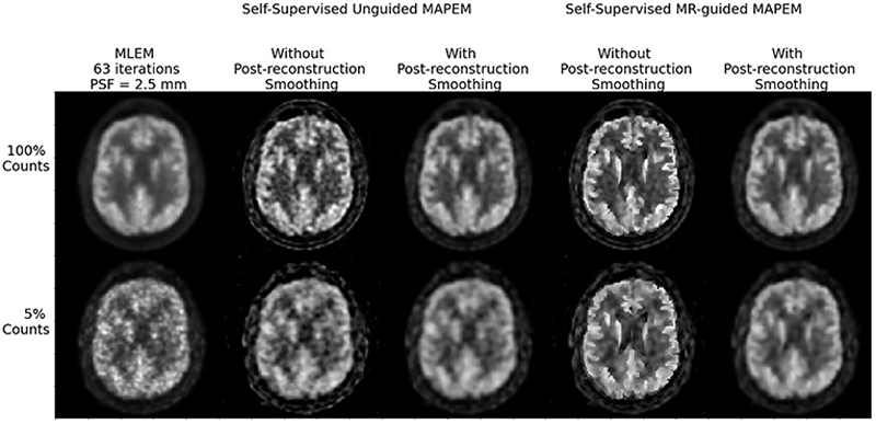
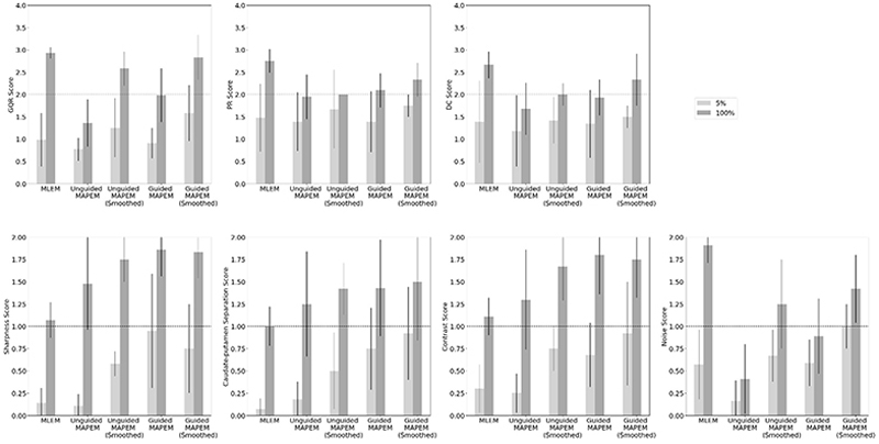
减少剂量正电子发射断层扫描(PET)降低了对患者的辐射剂量,降低了成本。然而,较低的计数数据会降低重建图像的质量。先进的重建方法有助于减轻图像质量损失,但重要的是从临床角度评估产生的图像。两位经验丰富的临床医生评估了[18F]FDG脑数据的四种PET重建算法,与临床标准参考(最大似然期望最大化(MLEM))进行比较,基于七个临床图像质量指标:整体质量评级、模式识别、诊断置信度(均在0-4之间)、清晰度、尾核-壳核分离、噪声和对比度(在0-2之间)。评估的重建方法是自我监督最大后验EM (MAPEM)的引导和非引导版本(其中引导案例使用患者的MR图像来控制平滑度惩罚)。对于重建的11个患者数据集中的3个,还考虑了MAPEM重建的后平滑版本,其中平滑使用分辨率建模中使用的点扩展函数。与MLEM相比,自我监督mr引导的MAPEM在清晰度、尾核-壳核分离和对比度方面观察到统计学上显著的改善。例如,MLEM在清晰度、尾壳核分离和对比度方面的得分为1-1.1分(满分为2分),而自我监督磁共振引导的MAPEM得分为1.5-1.75分。除了临床评估,预训练的卷积神经网络(cnn)被用来评估另外62张图像的图像质量。cnn向临床医生展示了类似的趋势,显示了它们作为自动独立观察者的潜力。临床和CNN评估均表明,当仅使用标准注射剂量的5%时,自我监督mr引导的MAPEM重建的整体性能与100% MLEM病例相匹配。这使得图像在临床上比标准的MLEM更有用。
本文章由计算机程序翻译,如有差异,请以英文原文为准。

Clinical and Deep-Learned Evaluation of MR-Guided Self-Supervised PET Reconstruction

Clinical and Deep-Learned Evaluation of MR-Guided Self-Supervised PET Reconstruction

Clinical and Deep-Learned Evaluation of MR-Guided Self-Supervised PET Reconstruction

Clinical and Deep-Learned Evaluation of MR-Guided Self-Supervised PET Reconstruction
Reduced dose positron emission tomography (PET) lowers the radiation dose to patients and reduces costs. Lower-count data, however, degrades reconstructed image quality. Advanced reconstruction methods help mitigate image quality losses, but it is important to assess the resulting images from a clinical perspective. Two experienced clinicians assessed four PET reconstruction algorithms for [18F]FDG brain data, compared to a clinical standard reference (maximum-likelihood expectation-maximization (MLEM)), based on seven clinical image quality metrics: global quality rating, pattern recognition, diagnostic confidence (all on a scale of 0–4), sharpness, caudate-putamen separation (CP), noise, and contrast (on a scale between 0–2). The reconstruction methods assessed were a guided and unguided version of self-supervised maximum a posteriori EM (MAPEM) (where the guidance case used the patient’s MR image to control the smoothness penalty). For 3 of the 11 patient datasets reconstructed, post-smoothed versions of the MAPEM reconstruction were also considered, where the smoothing was with the point-spread-function used in the resolution modelling. Statistically significant improvements were observed in sharpness, CP, and contrast for self-supervised MR-guided MAPEM compared to MLEM. For example, MLEM scored between 1-1.1 out of 2 for sharpness, CP, and contrast, whereas self-supervised MR-guided MAPEM scored between 1.5-1.75. In addition to the clinical evaluation, pretrained convolutional neural networks (CNNs) were used to assess the image quality of a further 62 images. The CNNs demonstrated similar trends to the clinician, showing their potential as automated standalone observers. Both the clinical and CNN assessments suggest when using only 5% of the standard injected dose, self-supervised MR-guided MAPEM reconstruction matches the 100% MLEM case for overall performance. This makes the images far more clinically useful than standard MLEM.
求助全文
通过发布文献求助,成功后即可免费获取论文全文。 去求助
来源期刊
IEEE Transactions on Radiation and Plasma Medical Sciences
IEEE Transactions on Radiation and Plasma Medical Sciences RADIOLOGY, NUCLEAR MEDICINE & MEDICAL IMAGING-
CiteScore
8.00
自引率
18.20%
发文量
109
×
引用
GB/T 7714-2015
复制
MLA
复制
APA
复制
导出至
BibTeX EndNote RefMan NoteFirst NoteExpress
×
提示
您的信息不完整,为了账户安全,请先补充。
现在去补充
×
提示
您因"违规操作"
具体请查看互助需知
我知道了
×
提示
确定
请完成安全验证×
copy
已复制链接
快去分享给好友吧!
我知道了
右上角分享
点击右上角分享
0
联系我们:info@booksci.cn Book学术提供免费学术资源搜索服务,方便国内外学者检索中英文文献。致力于提供最便捷和优质的服务体验。 Copyright © 2023 布克学术 All rights reserved.
京ICP备2023020795号-1
ghs 京公网安备 11010802042870号
Book学术文献互助
Book学术文献互助群
群 号:604180095
Book学术官方微信