Kyung Eun Lee, Sung Eun Song, Kyu Ran Cho, Min Sun Bae, Bo Kyoung Seo, Soo-Yeon Kim, Ok Hee Woo
{"title":"基于数字乳腺x线摄影的人工智能计算机辅助诊断在数字乳腺断层合成x线摄影中的表现。","authors":"Kyung Eun Lee, Sung Eun Song, Kyu Ran Cho, Min Sun Bae, Bo Kyoung Seo, Soo-Yeon Kim, Ok Hee Woo","doi":"10.3348/kjr.2024.0664","DOIUrl":null,"url":null,"abstract":"<p><strong>Objective: </strong>To test the performance of an artificial intelligence-based computer-aided diagnosis (AI-CAD) designed for full-field digital mammography (FFDM) when applied to synthetic mammography (SM).</p><p><strong>Materials and methods: </strong>We analyzed 501 women (mean age, 57 ± 11 years) who underwent preoperative mammography and breast cancer surgery. This cohort consisted of 1002 breasts, comprising 517 with cancer and 485 without. All patients underwent digital breast tomosynthesis (DBT) and FFDM during the preoperative workup. The SM is routinely reconstructed using DBT. Commercial AI-CAD (Lunit Insight MMG, version 1.1.7.2) was retrospectively applied to SM and FFDM to calculate the abnormality scores for each breast. The median abnormality scores were compared for the 517 breasts with cancer using the Wilcoxon signed-rank test. Calibration curves of abnormality scores were evaluated. The discrimination performance was analyzed using the area under the receiver operating characteristic curve (AUC), sensitivity, and specificity using a 10% preset threshold. Sensitivity and specificity were further analyzed according to the mammographic and pathological characteristics. The results of SM and FFDM were compared.</p><p><strong>Results: </strong>AI-CAD demonstrated a significantly lower median abnormality score (71% vs. 96%, <i>P</i> < 0.001) and poorer calibration performance for SM than for FFDM. SM exhibited lower sensitivity (76.2% vs. 82.8%, <i>P</i> < 0.001), higher specificity (95.5% vs. 91.8%, <i>P</i> < 0.001), and comparable AUC (0.86 vs. 0.87, <i>P</i> = 0.127) than FFDM. SM showed lower sensitivity than FFDM in asymptomatic breasts, dense breasts, ductal carcinoma in situ, T1, N0, and hormone receptor-positive/human epidermal growth factor receptor 2-negative cancers but showed higher specificity in non-cancerous dense breasts.</p><p><strong>Conclusion: </strong>AI-CAD showed lower abnormality scores and reduced calibration performance for SM than for FFDM. Furthermore, the 10% preset threshold resulted in different discrimination performances for the SM. Given these limitations, off-label application of the current AI-CAD to SM should be avoided.</p>","PeriodicalId":17881,"journal":{"name":"Korean Journal of Radiology","volume":"26 3","pages":"217-229"},"PeriodicalIF":5.3000,"publicationDate":"2025-03-01","publicationTypes":"Journal Article","fieldsOfStudy":null,"isOpenAccess":false,"openAccessPdf":"https://www.ncbi.nlm.nih.gov/pmc/articles/PMC11865904/pdf/","citationCount":"0","resultStr":"{\"title\":\"Performance of Digital Mammography-Based Artificial Intelligence Computer-Aided Diagnosis on Synthetic Mammography From Digital Breast Tomosynthesis.\",\"authors\":\"Kyung Eun Lee, Sung Eun Song, Kyu Ran Cho, Min Sun Bae, Bo Kyoung Seo, Soo-Yeon Kim, Ok Hee Woo\",\"doi\":\"10.3348/kjr.2024.0664\",\"DOIUrl\":null,\"url\":null,\"abstract\":\"<p><strong>Objective: </strong>To test the performance of an artificial intelligence-based computer-aided diagnosis (AI-CAD) designed for full-field digital mammography (FFDM) when applied to synthetic mammography (SM).</p><p><strong>Materials and methods: </strong>We analyzed 501 women (mean age, 57 ± 11 years) who underwent preoperative mammography and breast cancer surgery. This cohort consisted of 1002 breasts, comprising 517 with cancer and 485 without. All patients underwent digital breast tomosynthesis (DBT) and FFDM during the preoperative workup. The SM is routinely reconstructed using DBT. Commercial AI-CAD (Lunit Insight MMG, version 1.1.7.2) was retrospectively applied to SM and FFDM to calculate the abnormality scores for each breast. The median abnormality scores were compared for the 517 breasts with cancer using the Wilcoxon signed-rank test. Calibration curves of abnormality scores were evaluated. The discrimination performance was analyzed using the area under the receiver operating characteristic curve (AUC), sensitivity, and specificity using a 10% preset threshold. Sensitivity and specificity were further analyzed according to the mammographic and pathological characteristics. The results of SM and FFDM were compared.</p><p><strong>Results: </strong>AI-CAD demonstrated a significantly lower median abnormality score (71% vs. 96%, <i>P</i> < 0.001) and poorer calibration performance for SM than for FFDM. SM exhibited lower sensitivity (76.2% vs. 82.8%, <i>P</i> < 0.001), higher specificity (95.5% vs. 91.8%, <i>P</i> < 0.001), and comparable AUC (0.86 vs. 0.87, <i>P</i> = 0.127) than FFDM. SM showed lower sensitivity than FFDM in asymptomatic breasts, dense breasts, ductal carcinoma in situ, T1, N0, and hormone receptor-positive/human epidermal growth factor receptor 2-negative cancers but showed higher specificity in non-cancerous dense breasts.</p><p><strong>Conclusion: </strong>AI-CAD showed lower abnormality scores and reduced calibration performance for SM than for FFDM. Furthermore, the 10% preset threshold resulted in different discrimination performances for the SM. Given these limitations, off-label application of the current AI-CAD to SM should be avoided.</p>\",\"PeriodicalId\":17881,\"journal\":{\"name\":\"Korean Journal of Radiology\",\"volume\":\"26 3\",\"pages\":\"217-229\"},\"PeriodicalIF\":5.3000,\"publicationDate\":\"2025-03-01\",\"publicationTypes\":\"Journal Article\",\"fieldsOfStudy\":null,\"isOpenAccess\":false,\"openAccessPdf\":\"https://www.ncbi.nlm.nih.gov/pmc/articles/PMC11865904/pdf/\",\"citationCount\":\"0\",\"resultStr\":null,\"platform\":\"Semanticscholar\",\"paperid\":null,\"PeriodicalName\":\"Korean Journal of Radiology\",\"FirstCategoryId\":\"3\",\"ListUrlMain\":\"https://doi.org/10.3348/kjr.2024.0664\",\"RegionNum\":2,\"RegionCategory\":\"医学\",\"ArticlePicture\":[],\"TitleCN\":null,\"AbstractTextCN\":null,\"PMCID\":null,\"EPubDate\":\"\",\"PubModel\":\"\",\"JCR\":\"Q1\",\"JCRName\":\"RADIOLOGY, NUCLEAR MEDICINE & MEDICAL IMAGING\",\"Score\":null,\"Total\":0}","platform":"Semanticscholar","paperid":null,"PeriodicalName":"Korean Journal of Radiology","FirstCategoryId":"3","ListUrlMain":"https://doi.org/10.3348/kjr.2024.0664","RegionNum":2,"RegionCategory":"医学","ArticlePicture":[],"TitleCN":null,"AbstractTextCN":null,"PMCID":null,"EPubDate":"","PubModel":"","JCR":"Q1","JCRName":"RADIOLOGY, NUCLEAR MEDICINE & MEDICAL IMAGING","Score":null,"Total":0}
引用次数: 0
摘要
目的:研究基于人工智能的全视场数字乳房x线摄影(FFDM)计算机辅助诊断(AI-CAD)在合成乳房x线摄影(SM)中的应用效果。材料和方法:我们分析了501例(平均年龄57±11岁)行术前乳房x光检查和乳腺癌手术的妇女。该队列包括1002个乳房,其中517个患有癌症,485个没有。所有患者术前均行数字乳腺断层合成(DBT)和FFDM。常规使用DBT重建SM。将商业AI-CAD (Lunit Insight MMG,版本1.1.7.2)回顾性应用于SM和FFDM,计算每个乳房的异常评分。使用Wilcoxon符号秩检验比较517例乳腺癌的中位异常评分。评估异常评分的校正曲线。使用受试者工作特征曲线下面积(AUC)、灵敏度和特异性(10%预设阈值)分析识别性能。根据x线摄影和病理特征进一步分析敏感性和特异性。比较SM和FFDM的结果。结果:与FFDM相比,AI-CAD显示出明显较低的异常评分中位数(71%对96%,P < 0.001), SM的校准性能较差。SM的敏感性较低(76.2% vs. 82.8%, P < 0.001),特异性较高(95.5% vs. 91.8%, P < 0.001), AUC与FFDM相当(0.86 vs. 0.87, P = 0.127)。SM在无症状乳房、致密乳房、导管原位癌、T1、N0和激素受体阳性/人表皮生长因子受体2阴性肿瘤中的敏感性低于FFDM,但在非癌性致密乳房中表现出更高的特异性。结论:与FFDM相比,AI-CAD对SM的异常评分较低,校正效果较差。此外,10%的预设阈值对SM的识别性能也有不同的影响。鉴于这些限制,应避免将当前的AI-CAD在标签外应用于SM。
Performance of Digital Mammography-Based Artificial Intelligence Computer-Aided Diagnosis on Synthetic Mammography From Digital Breast Tomosynthesis.
Objective: To test the performance of an artificial intelligence-based computer-aided diagnosis (AI-CAD) designed for full-field digital mammography (FFDM) when applied to synthetic mammography (SM).
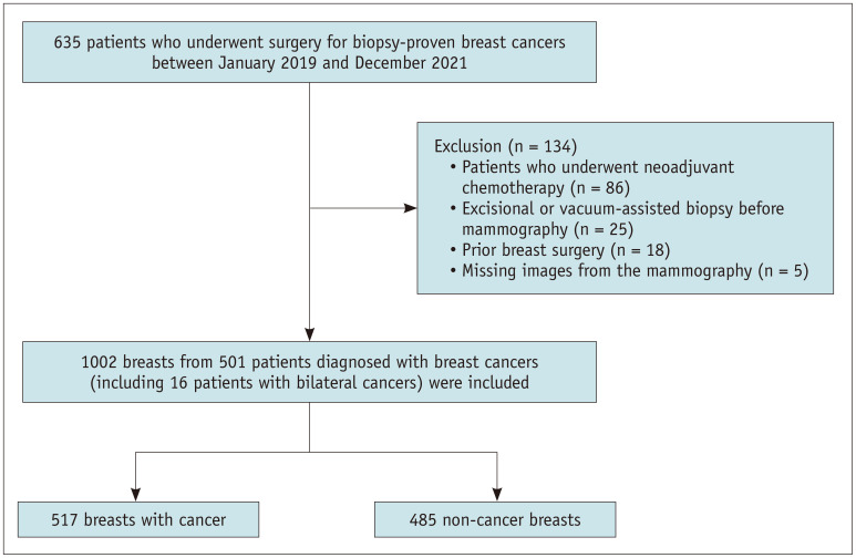
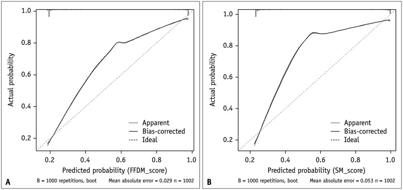
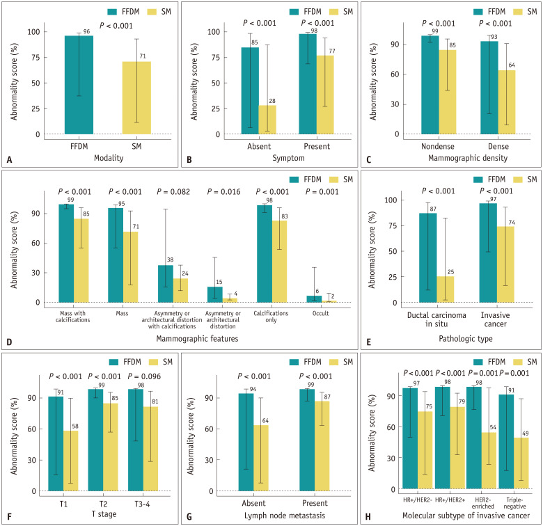
Materials and methods: We analyzed 501 women (mean age, 57 ± 11 years) who underwent preoperative mammography and breast cancer surgery. This cohort consisted of 1002 breasts, comprising 517 with cancer and 485 without. All patients underwent digital breast tomosynthesis (DBT) and FFDM during the preoperative workup. The SM is routinely reconstructed using DBT. Commercial AI-CAD (Lunit Insight MMG, version 1.1.7.2) was retrospectively applied to SM and FFDM to calculate the abnormality scores for each breast. The median abnormality scores were compared for the 517 breasts with cancer using the Wilcoxon signed-rank test. Calibration curves of abnormality scores were evaluated. The discrimination performance was analyzed using the area under the receiver operating characteristic curve (AUC), sensitivity, and specificity using a 10% preset threshold. Sensitivity and specificity were further analyzed according to the mammographic and pathological characteristics. The results of SM and FFDM were compared.
Results: AI-CAD demonstrated a significantly lower median abnormality score (71% vs. 96%, P < 0.001) and poorer calibration performance for SM than for FFDM. SM exhibited lower sensitivity (76.2% vs. 82.8%, P < 0.001), higher specificity (95.5% vs. 91.8%, P < 0.001), and comparable AUC (0.86 vs. 0.87, P = 0.127) than FFDM. SM showed lower sensitivity than FFDM in asymptomatic breasts, dense breasts, ductal carcinoma in situ, T1, N0, and hormone receptor-positive/human epidermal growth factor receptor 2-negative cancers but showed higher specificity in non-cancerous dense breasts.
Conclusion: AI-CAD showed lower abnormality scores and reduced calibration performance for SM than for FFDM. Furthermore, the 10% preset threshold resulted in different discrimination performances for the SM. Given these limitations, off-label application of the current AI-CAD to SM should be avoided.
期刊介绍:
The inaugural issue of the Korean J Radiol came out in March 2000. Our journal aims to produce and propagate knowledge on radiologic imaging and related sciences.
A unique feature of the articles published in the Journal will be their reflection of global trends in radiology combined with an East-Asian perspective. Geographic differences in disease prevalence will be reflected in the contents of papers, and this will serve to enrich our body of knowledge.
World''s outstanding radiologists from many countries are serving as editorial board of our journal.

 求助内容:
求助内容: 应助结果提醒方式:


