Samuel Guay, Camille Charlebois-Plante, Sophie-Andrée Vinet, Marie-Eve Bourassa, Louis De Beaumont
{"title":"持续性脑震荡后综合征运动员的结构磁共振成像脑年龄调查。","authors":"Samuel Guay, Camille Charlebois-Plante, Sophie-Andrée Vinet, Marie-Eve Bourassa, Louis De Beaumont","doi":"10.1089/neur.2024.0094","DOIUrl":null,"url":null,"abstract":"<p><p>Brain age prediction algorithms using structural magnetic resonance imaging (MRI) estimate the biological age of the brain by comparing it to a normal aging trajectory, allowing for the identification of deviations that may indicate slower or accelerated biological aging. Traumatic brain injury (TBI) and sports-related concussion (SRC) have been associated with greater brain age gap (BAG) compared to healthy controls. In this study, we aimed to investigate BAG in athletes suffering from persistent postconcussion syndrome (PCS+) compared to PCS- athletes, and used SHapley Additive exPlanations (SHAP), an explainable artificial intelligence framework, to provide further details on which specific features drive the brain age predictions. Brain age was derived from T1-weighted MRI images in a cohort of 50 athletes (24 with PCS+) from 22 to 73 years old from the general population. The results revealed that athletes with PCS+ had a brain age approximately 5 years older than the PCS- athletes, with no clinical variable associated with it. Exploratory analyses also showed a greater brain age in athletes who self-reported five or more SRCs. Regarding SHAP, the third ventricle was found to be the most informative feature in the PCS+ group, while the superior temporal sulcus posterior area was more informative in the PCS- group. This study demonstrated the potential of using brain age and explainable artificial intelligence frameworks to study athletes with PCS. Further research is needed to explore the underlying mechanisms driving brain aging in this population and to identify potential biomarkers for early detection and intervention.</p>","PeriodicalId":74300,"journal":{"name":"Neurotrauma reports","volume":"6 1","pages":"136-147"},"PeriodicalIF":1.8000,"publicationDate":"2025-01-31","publicationTypes":"Journal Article","fieldsOfStudy":null,"isOpenAccess":false,"openAccessPdf":"https://www.ncbi.nlm.nih.gov/pmc/articles/PMC11839523/pdf/","citationCount":"0","resultStr":"{\"title\":\"Structural Magnetic Resonance Imaging Brain Age Investigation in Athletes with Persistent Postconcussion Syndrome.\",\"authors\":\"Samuel Guay, Camille Charlebois-Plante, Sophie-Andrée Vinet, Marie-Eve Bourassa, Louis De Beaumont\",\"doi\":\"10.1089/neur.2024.0094\",\"DOIUrl\":null,\"url\":null,\"abstract\":\"<p><p>Brain age prediction algorithms using structural magnetic resonance imaging (MRI) estimate the biological age of the brain by comparing it to a normal aging trajectory, allowing for the identification of deviations that may indicate slower or accelerated biological aging. Traumatic brain injury (TBI) and sports-related concussion (SRC) have been associated with greater brain age gap (BAG) compared to healthy controls. In this study, we aimed to investigate BAG in athletes suffering from persistent postconcussion syndrome (PCS+) compared to PCS- athletes, and used SHapley Additive exPlanations (SHAP), an explainable artificial intelligence framework, to provide further details on which specific features drive the brain age predictions. Brain age was derived from T1-weighted MRI images in a cohort of 50 athletes (24 with PCS+) from 22 to 73 years old from the general population. The results revealed that athletes with PCS+ had a brain age approximately 5 years older than the PCS- athletes, with no clinical variable associated with it. Exploratory analyses also showed a greater brain age in athletes who self-reported five or more SRCs. Regarding SHAP, the third ventricle was found to be the most informative feature in the PCS+ group, while the superior temporal sulcus posterior area was more informative in the PCS- group. This study demonstrated the potential of using brain age and explainable artificial intelligence frameworks to study athletes with PCS. Further research is needed to explore the underlying mechanisms driving brain aging in this population and to identify potential biomarkers for early detection and intervention.</p>\",\"PeriodicalId\":74300,\"journal\":{\"name\":\"Neurotrauma reports\",\"volume\":\"6 1\",\"pages\":\"136-147\"},\"PeriodicalIF\":1.8000,\"publicationDate\":\"2025-01-31\",\"publicationTypes\":\"Journal Article\",\"fieldsOfStudy\":null,\"isOpenAccess\":false,\"openAccessPdf\":\"https://www.ncbi.nlm.nih.gov/pmc/articles/PMC11839523/pdf/\",\"citationCount\":\"0\",\"resultStr\":null,\"platform\":\"Semanticscholar\",\"paperid\":null,\"PeriodicalName\":\"Neurotrauma reports\",\"FirstCategoryId\":\"1085\",\"ListUrlMain\":\"https://doi.org/10.1089/neur.2024.0094\",\"RegionNum\":0,\"RegionCategory\":null,\"ArticlePicture\":[],\"TitleCN\":null,\"AbstractTextCN\":null,\"PMCID\":null,\"EPubDate\":\"2025/1/1 0:00:00\",\"PubModel\":\"eCollection\",\"JCR\":\"Q3\",\"JCRName\":\"CLINICAL NEUROLOGY\",\"Score\":null,\"Total\":0}","platform":"Semanticscholar","paperid":null,"PeriodicalName":"Neurotrauma reports","FirstCategoryId":"1085","ListUrlMain":"https://doi.org/10.1089/neur.2024.0094","RegionNum":0,"RegionCategory":null,"ArticlePicture":[],"TitleCN":null,"AbstractTextCN":null,"PMCID":null,"EPubDate":"2025/1/1 0:00:00","PubModel":"eCollection","JCR":"Q3","JCRName":"CLINICAL NEUROLOGY","Score":null,"Total":0}
Structural Magnetic Resonance Imaging Brain Age Investigation in Athletes with Persistent Postconcussion Syndrome.
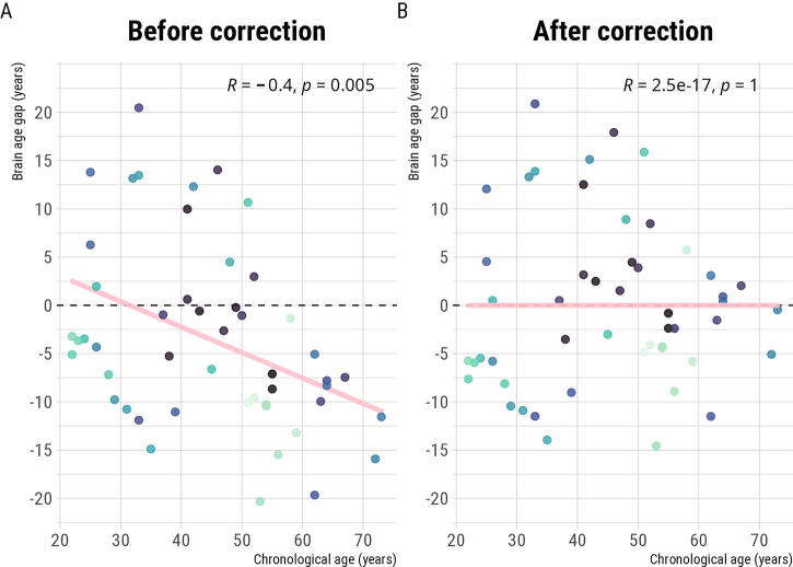
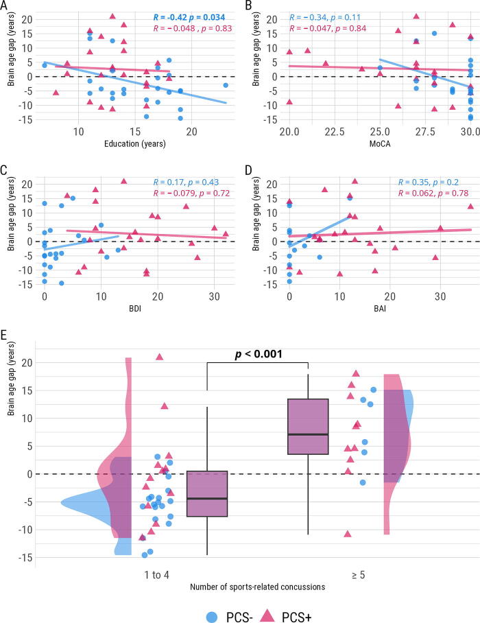
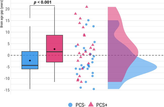
Brain age prediction algorithms using structural magnetic resonance imaging (MRI) estimate the biological age of the brain by comparing it to a normal aging trajectory, allowing for the identification of deviations that may indicate slower or accelerated biological aging. Traumatic brain injury (TBI) and sports-related concussion (SRC) have been associated with greater brain age gap (BAG) compared to healthy controls. In this study, we aimed to investigate BAG in athletes suffering from persistent postconcussion syndrome (PCS+) compared to PCS- athletes, and used SHapley Additive exPlanations (SHAP), an explainable artificial intelligence framework, to provide further details on which specific features drive the brain age predictions. Brain age was derived from T1-weighted MRI images in a cohort of 50 athletes (24 with PCS+) from 22 to 73 years old from the general population. The results revealed that athletes with PCS+ had a brain age approximately 5 years older than the PCS- athletes, with no clinical variable associated with it. Exploratory analyses also showed a greater brain age in athletes who self-reported five or more SRCs. Regarding SHAP, the third ventricle was found to be the most informative feature in the PCS+ group, while the superior temporal sulcus posterior area was more informative in the PCS- group. This study demonstrated the potential of using brain age and explainable artificial intelligence frameworks to study athletes with PCS. Further research is needed to explore the underlying mechanisms driving brain aging in this population and to identify potential biomarkers for early detection and intervention.

 求助内容:
求助内容: 应助结果提醒方式:


