{"title":"应用新型腹腔镜荧光光谱系统及近红外荧光夹进行腹腔镜胃癌切除术1例。","authors":"Shion Uemura, Yuma Ebihara, Kazuya Konishi, Satoshi Hirano","doi":"10.70352/scrj.cr.24-0028","DOIUrl":null,"url":null,"abstract":"<p><strong>Introduction: </strong>In laparoscopic gastrectomy, accurate marking of the lesion site is essential in determining the resection line of the stomach, owing to the lack of haptics and the direct link between negative pathological margins and prognosis. Intraoperative endoscopy may require personnel and prolong the operation time, whereas preoperative endoscopic tattooing using India ink faces problems related to the spread of ink and visibility. ZEOCLIP FS (Zeon Medical, Tokyo, Japan) is a clip made of fluorescent resin, covered by insurance since March 2019. It can be visualized from the serosal side using a near-infrared scope; however, its weak fluorescence intensity often poses viewing difficulties. Lumifinder (ADVANTEST, Tokyo, Japan) is a laparoscopic fluorescence spectrum system available for clinical use since February 2023. It can measure fluorescence intensity using a near-infrared laser and detect weak fluorescent signals. We report a case of gastric cancer in which the location of the lesion was confirmed intraoperatively using ZEOCLIP FS and Lumifinder.</p><p><strong>Case presentation: </strong>A man in his 80s was diagnosed with gastric cancer following an examination for anemia. Two lesions were found: a 0-IIc type (cT1) at the lesser curvature of the gastric angle and a type 1 tumor (cT2) at the anterior wall of the upper gastric body. The preoperative assessment indicated no lymph node or distant metastasis. The tumor was diagnosed as cStage I and laparoscopic distal gastrectomy was planned. Two ZEOCLIP FS clips were placed on the oral side of the tumor on the anterior wall of the upper gastric body on the day before surgery. During surgery, fluorescent signals from the clips were detected using Lumifinder, enabling easy confirmation of the lesion location and determination of the gastric resection line.</p><p><strong>Conclusions: </strong>The combined use of ZEOCLIP FS and Lumifinder was a useful new method for identifying the appropriate resection line of the stomach. We plan to evaluate this method further in additional cases to enhance the detection efficacy.</p>","PeriodicalId":22096,"journal":{"name":"Surgical Case Reports","volume":"11 1","pages":""},"PeriodicalIF":0.7000,"publicationDate":"2025-01-01","publicationTypes":"Journal Article","fieldsOfStudy":null,"isOpenAccess":false,"openAccessPdf":"https://www.ncbi.nlm.nih.gov/pmc/articles/PMC11842931/pdf/","citationCount":"0","resultStr":"{\"title\":\"A Case of Laparoscopic Resection of Gastric Cancer Using Novel Laparoscopic Fluorescence Spectrum System and Near-Infrared Fluorescent Clips.\",\"authors\":\"Shion Uemura, Yuma Ebihara, Kazuya Konishi, Satoshi Hirano\",\"doi\":\"10.70352/scrj.cr.24-0028\",\"DOIUrl\":null,\"url\":null,\"abstract\":\"<p><strong>Introduction: </strong>In laparoscopic gastrectomy, accurate marking of the lesion site is essential in determining the resection line of the stomach, owing to the lack of haptics and the direct link between negative pathological margins and prognosis. Intraoperative endoscopy may require personnel and prolong the operation time, whereas preoperative endoscopic tattooing using India ink faces problems related to the spread of ink and visibility. ZEOCLIP FS (Zeon Medical, Tokyo, Japan) is a clip made of fluorescent resin, covered by insurance since March 2019. It can be visualized from the serosal side using a near-infrared scope; however, its weak fluorescence intensity often poses viewing difficulties. Lumifinder (ADVANTEST, Tokyo, Japan) is a laparoscopic fluorescence spectrum system available for clinical use since February 2023. It can measure fluorescence intensity using a near-infrared laser and detect weak fluorescent signals. We report a case of gastric cancer in which the location of the lesion was confirmed intraoperatively using ZEOCLIP FS and Lumifinder.</p><p><strong>Case presentation: </strong>A man in his 80s was diagnosed with gastric cancer following an examination for anemia. Two lesions were found: a 0-IIc type (cT1) at the lesser curvature of the gastric angle and a type 1 tumor (cT2) at the anterior wall of the upper gastric body. The preoperative assessment indicated no lymph node or distant metastasis. The tumor was diagnosed as cStage I and laparoscopic distal gastrectomy was planned. Two ZEOCLIP FS clips were placed on the oral side of the tumor on the anterior wall of the upper gastric body on the day before surgery. During surgery, fluorescent signals from the clips were detected using Lumifinder, enabling easy confirmation of the lesion location and determination of the gastric resection line.</p><p><strong>Conclusions: </strong>The combined use of ZEOCLIP FS and Lumifinder was a useful new method for identifying the appropriate resection line of the stomach. We plan to evaluate this method further in additional cases to enhance the detection efficacy.</p>\",\"PeriodicalId\":22096,\"journal\":{\"name\":\"Surgical Case Reports\",\"volume\":\"11 1\",\"pages\":\"\"},\"PeriodicalIF\":0.7000,\"publicationDate\":\"2025-01-01\",\"publicationTypes\":\"Journal Article\",\"fieldsOfStudy\":null,\"isOpenAccess\":false,\"openAccessPdf\":\"https://www.ncbi.nlm.nih.gov/pmc/articles/PMC11842931/pdf/\",\"citationCount\":\"0\",\"resultStr\":null,\"platform\":\"Semanticscholar\",\"paperid\":null,\"PeriodicalName\":\"Surgical Case Reports\",\"FirstCategoryId\":\"1085\",\"ListUrlMain\":\"https://doi.org/10.70352/scrj.cr.24-0028\",\"RegionNum\":0,\"RegionCategory\":null,\"ArticlePicture\":[],\"TitleCN\":null,\"AbstractTextCN\":null,\"PMCID\":null,\"EPubDate\":\"2025/2/1 0:00:00\",\"PubModel\":\"Epub\",\"JCR\":\"Q4\",\"JCRName\":\"SURGERY\",\"Score\":null,\"Total\":0}","platform":"Semanticscholar","paperid":null,"PeriodicalName":"Surgical Case Reports","FirstCategoryId":"1085","ListUrlMain":"https://doi.org/10.70352/scrj.cr.24-0028","RegionNum":0,"RegionCategory":null,"ArticlePicture":[],"TitleCN":null,"AbstractTextCN":null,"PMCID":null,"EPubDate":"2025/2/1 0:00:00","PubModel":"Epub","JCR":"Q4","JCRName":"SURGERY","Score":null,"Total":0}
引用次数: 0
摘要
导论:在腹腔镜胃切除术中,由于缺乏触觉,病理边缘阴性与预后直接相关,准确标记病变部位对于确定胃的切除线至关重要。术中内窥镜检查可能需要人员和延长手术时间,而术前使用印度墨水进行内窥镜纹身则面临墨水扩散和可见性等问题。ZEOCLIP FS (Zeon Medical, Tokyo, Japan)是一种荧光树脂制成的夹子,自2019年3月起投保。它可以使用近红外范围从浆膜侧可视化;然而,其微弱的荧光强度往往造成观察困难。Lumifinder (ADVANTEST, Tokyo, Japan)是一种腹腔镜荧光光谱系统,自2023年2月起可用于临床。它可以使用近红外激光测量荧光强度,并检测微弱的荧光信号。我们报告一例胃癌,术中使用ZEOCLIP FS和Lumifinder确认病变位置。病例介绍:一名80多岁的男子在贫血检查后被诊断为胃癌。发现两个病变:胃角小弯0-IIc型(cT1)和胃上体前壁1型肿瘤(cT2)。术前评估显示无淋巴结或远处转移。肿瘤诊断为c期I期,计划行腹腔镜远端胃切除术。术前1天将2个ZEOCLIP FS夹放置于肿瘤口腔侧胃上体前壁。手术过程中,使用Lumifinder检测夹子发出的荧光信号,方便确认病变位置和确定胃切除线。结论:ZEOCLIP FS与Lumifinder联合使用是一种确定胃合适切除线的有效新方法。我们计划在其他病例中进一步评估该方法,以提高检测效率。
A Case of Laparoscopic Resection of Gastric Cancer Using Novel Laparoscopic Fluorescence Spectrum System and Near-Infrared Fluorescent Clips.
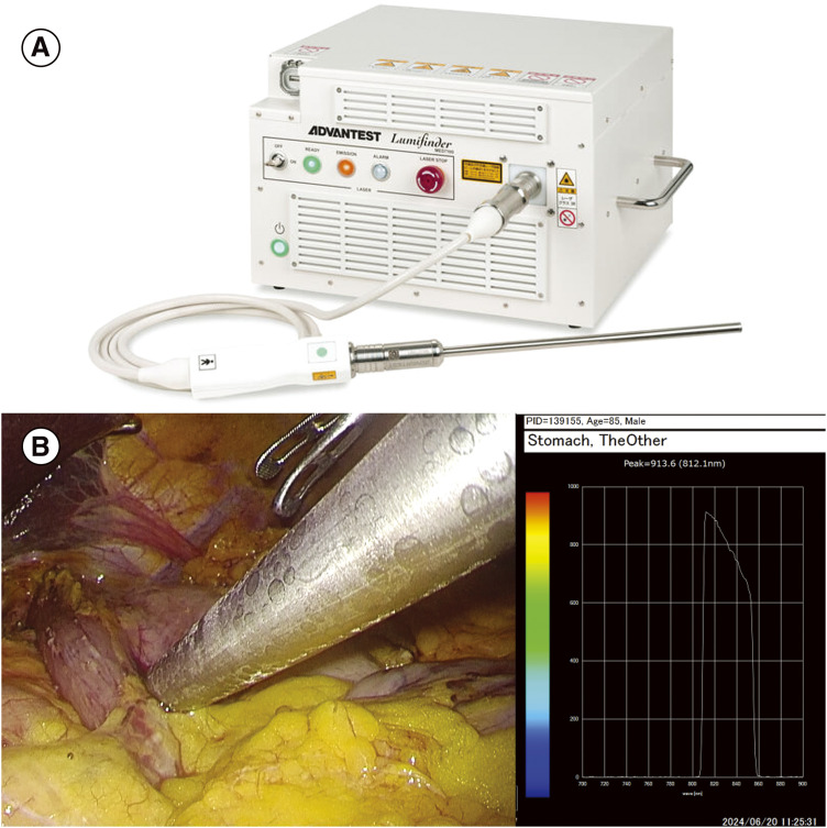
Introduction: In laparoscopic gastrectomy, accurate marking of the lesion site is essential in determining the resection line of the stomach, owing to the lack of haptics and the direct link between negative pathological margins and prognosis. Intraoperative endoscopy may require personnel and prolong the operation time, whereas preoperative endoscopic tattooing using India ink faces problems related to the spread of ink and visibility. ZEOCLIP FS (Zeon Medical, Tokyo, Japan) is a clip made of fluorescent resin, covered by insurance since March 2019. It can be visualized from the serosal side using a near-infrared scope; however, its weak fluorescence intensity often poses viewing difficulties. Lumifinder (ADVANTEST, Tokyo, Japan) is a laparoscopic fluorescence spectrum system available for clinical use since February 2023. It can measure fluorescence intensity using a near-infrared laser and detect weak fluorescent signals. We report a case of gastric cancer in which the location of the lesion was confirmed intraoperatively using ZEOCLIP FS and Lumifinder.
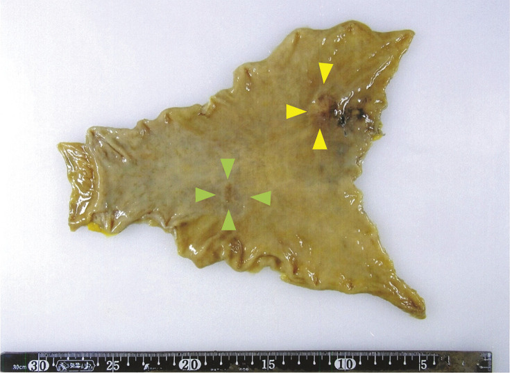
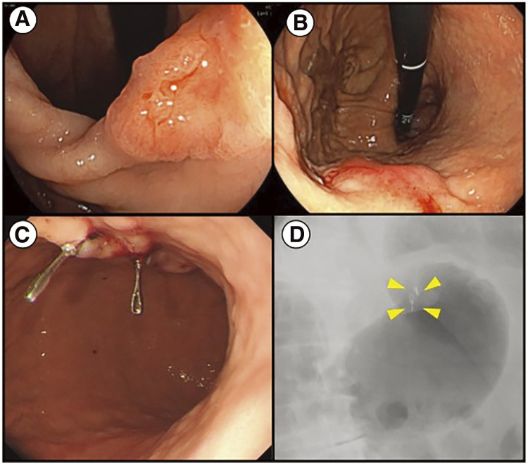
Case presentation: A man in his 80s was diagnosed with gastric cancer following an examination for anemia. Two lesions were found: a 0-IIc type (cT1) at the lesser curvature of the gastric angle and a type 1 tumor (cT2) at the anterior wall of the upper gastric body. The preoperative assessment indicated no lymph node or distant metastasis. The tumor was diagnosed as cStage I and laparoscopic distal gastrectomy was planned. Two ZEOCLIP FS clips were placed on the oral side of the tumor on the anterior wall of the upper gastric body on the day before surgery. During surgery, fluorescent signals from the clips were detected using Lumifinder, enabling easy confirmation of the lesion location and determination of the gastric resection line.
Conclusions: The combined use of ZEOCLIP FS and Lumifinder was a useful new method for identifying the appropriate resection line of the stomach. We plan to evaluate this method further in additional cases to enhance the detection efficacy.

 求助内容:
求助内容: 应助结果提醒方式:


