Selene Barone, Francesco Bennardo, Marianna Salviati, Alessandro Antonelli, Amerigo Giudice
{"title":"评估富血小板纤维蛋白(PRF)在下颌第三磨牙手术中的实用性与3D面部肿胀分析:一项裂口随机临床试验。","authors":"Selene Barone, Francesco Bennardo, Marianna Salviati, Alessandro Antonelli, Amerigo Giudice","doi":"10.1186/s13005-025-00482-0","DOIUrl":null,"url":null,"abstract":"<p><strong>Background: </strong>Third molar surgery is associated with various postoperative complications (PC). Different strategies, including the application of platelet-rich fibrin (PRF), have been implemented to reduce PC. Digital technologies have proven useful in objectively assessing postoperative facial swelling. This study aimed to evaluate the effect of PRF on reducing facial swelling after lower third molar surgery using a 3D face scanner.</p><p><strong>Methods: </strong>A randomized split-mouth clinical trial was set up and 32 patients (18 to 32 years), requiring extraction of both mandibular third molars, were recruited at the Oral Surgery Clinic of the Magna Graecia University of Catanzaro. The primary predictive variable was the application or not of PRF plugs and membranes in the post-extraction socket. Primary outcome variable was facial swelling recorded with a face scanner preoperatively (T0), after three (T1) and seven (T2) days. Qualitative and quantitative data analysis were conducted following an automated and standardized imaging analysis workflow using the 3D Slicer software. Secondary outcome variables were trismus, recorded by measuring the maximum buccal opening with a caliper, pain, recorded using a visual analogue scale (VAS), and duration of the surgery. Descriptive and bivariate analysis were performed by setting the significance level [Formula: see text] = 0.05.</p><p><strong>Results: </strong>All patients exhibited a significant increase in facial swelling at T1, followed by a subsequent reduction from day 3 to day 7, with a slight persistence of edema observed on the seventh day. No significant data emerged from the statistical analysis conducted. Linear differences in PRF group reported improved values of postoperative swelling only in the T1-T2 and T0-T2 phases of analysis. Volumetric differences favored PRF group compared with control group in all phases. VAS was lower in PRF group only at T2, compared with control group.</p><p><strong>Conclusions: </strong>Application of PRF in post-extraction sockets showed effectiveness in reducing facial swelling. Its advantages, including accessibility, cost-effectiveness, and absence of adverse reactions, make it an optimal treatment choice in reducing post-surgical sequelae.</p>","PeriodicalId":12994,"journal":{"name":"Head & Face Medicine","volume":"21 1","pages":"8"},"PeriodicalIF":2.4000,"publicationDate":"2025-02-22","publicationTypes":"Journal Article","fieldsOfStudy":null,"isOpenAccess":false,"openAccessPdf":"https://www.ncbi.nlm.nih.gov/pmc/articles/PMC11846411/pdf/","citationCount":"0","resultStr":"{\"title\":\"Evaluation of the usefulness of platelet-rich fibrin (PRF) in mandibular third molar surgery with 3D facial swelling analysis: a split-mouth randomized clinical trial.\",\"authors\":\"Selene Barone, Francesco Bennardo, Marianna Salviati, Alessandro Antonelli, Amerigo Giudice\",\"doi\":\"10.1186/s13005-025-00482-0\",\"DOIUrl\":null,\"url\":null,\"abstract\":\"<p><strong>Background: </strong>Third molar surgery is associated with various postoperative complications (PC). Different strategies, including the application of platelet-rich fibrin (PRF), have been implemented to reduce PC. Digital technologies have proven useful in objectively assessing postoperative facial swelling. This study aimed to evaluate the effect of PRF on reducing facial swelling after lower third molar surgery using a 3D face scanner.</p><p><strong>Methods: </strong>A randomized split-mouth clinical trial was set up and 32 patients (18 to 32 years), requiring extraction of both mandibular third molars, were recruited at the Oral Surgery Clinic of the Magna Graecia University of Catanzaro. The primary predictive variable was the application or not of PRF plugs and membranes in the post-extraction socket. Primary outcome variable was facial swelling recorded with a face scanner preoperatively (T0), after three (T1) and seven (T2) days. Qualitative and quantitative data analysis were conducted following an automated and standardized imaging analysis workflow using the 3D Slicer software. Secondary outcome variables were trismus, recorded by measuring the maximum buccal opening with a caliper, pain, recorded using a visual analogue scale (VAS), and duration of the surgery. Descriptive and bivariate analysis were performed by setting the significance level [Formula: see text] = 0.05.</p><p><strong>Results: </strong>All patients exhibited a significant increase in facial swelling at T1, followed by a subsequent reduction from day 3 to day 7, with a slight persistence of edema observed on the seventh day. No significant data emerged from the statistical analysis conducted. Linear differences in PRF group reported improved values of postoperative swelling only in the T1-T2 and T0-T2 phases of analysis. Volumetric differences favored PRF group compared with control group in all phases. VAS was lower in PRF group only at T2, compared with control group.</p><p><strong>Conclusions: </strong>Application of PRF in post-extraction sockets showed effectiveness in reducing facial swelling. Its advantages, including accessibility, cost-effectiveness, and absence of adverse reactions, make it an optimal treatment choice in reducing post-surgical sequelae.</p>\",\"PeriodicalId\":12994,\"journal\":{\"name\":\"Head & Face Medicine\",\"volume\":\"21 1\",\"pages\":\"8\"},\"PeriodicalIF\":2.4000,\"publicationDate\":\"2025-02-22\",\"publicationTypes\":\"Journal Article\",\"fieldsOfStudy\":null,\"isOpenAccess\":false,\"openAccessPdf\":\"https://www.ncbi.nlm.nih.gov/pmc/articles/PMC11846411/pdf/\",\"citationCount\":\"0\",\"resultStr\":null,\"platform\":\"Semanticscholar\",\"paperid\":null,\"PeriodicalName\":\"Head & Face Medicine\",\"FirstCategoryId\":\"3\",\"ListUrlMain\":\"https://doi.org/10.1186/s13005-025-00482-0\",\"RegionNum\":2,\"RegionCategory\":\"医学\",\"ArticlePicture\":[],\"TitleCN\":null,\"AbstractTextCN\":null,\"PMCID\":null,\"EPubDate\":\"\",\"PubModel\":\"\",\"JCR\":\"Q2\",\"JCRName\":\"DENTISTRY, ORAL SURGERY & MEDICINE\",\"Score\":null,\"Total\":0}","platform":"Semanticscholar","paperid":null,"PeriodicalName":"Head & Face Medicine","FirstCategoryId":"3","ListUrlMain":"https://doi.org/10.1186/s13005-025-00482-0","RegionNum":2,"RegionCategory":"医学","ArticlePicture":[],"TitleCN":null,"AbstractTextCN":null,"PMCID":null,"EPubDate":"","PubModel":"","JCR":"Q2","JCRName":"DENTISTRY, ORAL SURGERY & MEDICINE","Score":null,"Total":0}
引用次数: 0
摘要
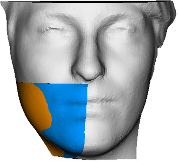
背景:第三磨牙手术与各种术后并发症(PC)相关。已经实施了不同的策略,包括应用富血小板纤维蛋白(PRF)来降低PC。数字技术在客观评估术后面部肿胀方面已被证明是有用的。本研究旨在利用3D面部扫描仪评估PRF对减少下第三磨牙手术后面部肿胀的影响。方法:在Catanzaro Magna Graecia大学口腔外科诊所建立随机裂口临床试验,招募32例(18 ~ 32岁)需要拔除下颌第三磨牙的患者。主要的预测变量是是否在拔牙后的窝中应用PRF塞和膜。主要结局变量是术前(T0)、术后3天(T1)和7天(T2)用面部扫描仪记录面部肿胀。定性和定量数据分析是根据使用3D切片器软件的自动化和标准化成像分析工作流程进行的。次要结果变量是牙关,用卡尺测量最大颊开口记录,疼痛,用视觉模拟量表(VAS)记录,以及手术持续时间。采用显著性水平(公式见文)= 0.05进行描述性和双变量分析。结果:所有患者在T1时面部肿胀明显增加,随后从第3天到第7天减轻,第7天观察到轻微的持续水肿。从所进行的统计分析中没有发现有意义的数据。PRF组的线性差异仅在分析的T1-T2和T0-T2阶段报告了术后肿胀的改善值。各期体积差异均有利于PRF组。PRF组仅在T2时VAS低于对照组。结论:PRF应用于拔牙后牙槽能有效减轻面部肿胀。其优点包括可及性、成本效益和无不良反应,使其成为减少术后后遗症的最佳治疗选择。
Evaluation of the usefulness of platelet-rich fibrin (PRF) in mandibular third molar surgery with 3D facial swelling analysis: a split-mouth randomized clinical trial.
Background: Third molar surgery is associated with various postoperative complications (PC). Different strategies, including the application of platelet-rich fibrin (PRF), have been implemented to reduce PC. Digital technologies have proven useful in objectively assessing postoperative facial swelling. This study aimed to evaluate the effect of PRF on reducing facial swelling after lower third molar surgery using a 3D face scanner.
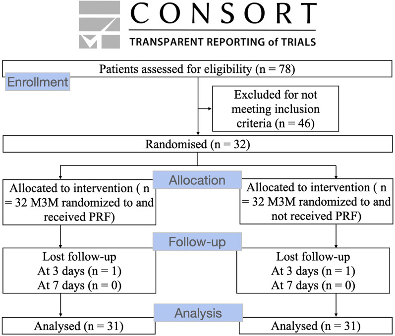
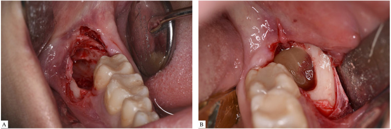
Methods: A randomized split-mouth clinical trial was set up and 32 patients (18 to 32 years), requiring extraction of both mandibular third molars, were recruited at the Oral Surgery Clinic of the Magna Graecia University of Catanzaro. The primary predictive variable was the application or not of PRF plugs and membranes in the post-extraction socket. Primary outcome variable was facial swelling recorded with a face scanner preoperatively (T0), after three (T1) and seven (T2) days. Qualitative and quantitative data analysis were conducted following an automated and standardized imaging analysis workflow using the 3D Slicer software. Secondary outcome variables were trismus, recorded by measuring the maximum buccal opening with a caliper, pain, recorded using a visual analogue scale (VAS), and duration of the surgery. Descriptive and bivariate analysis were performed by setting the significance level [Formula: see text] = 0.05.
Results: All patients exhibited a significant increase in facial swelling at T1, followed by a subsequent reduction from day 3 to day 7, with a slight persistence of edema observed on the seventh day. No significant data emerged from the statistical analysis conducted. Linear differences in PRF group reported improved values of postoperative swelling only in the T1-T2 and T0-T2 phases of analysis. Volumetric differences favored PRF group compared with control group in all phases. VAS was lower in PRF group only at T2, compared with control group.
Conclusions: Application of PRF in post-extraction sockets showed effectiveness in reducing facial swelling. Its advantages, including accessibility, cost-effectiveness, and absence of adverse reactions, make it an optimal treatment choice in reducing post-surgical sequelae.
期刊介绍:
Head & Face Medicine is a multidisciplinary open access journal that publishes basic and clinical research concerning all aspects of cranial, facial and oral conditions.
The journal covers all aspects of cranial, facial and oral diseases and their management. It has been designed as a multidisciplinary journal for clinicians and researchers involved in the diagnostic and therapeutic aspects of diseases which affect the human head and face. The journal is wide-ranging, covering the development, aetiology, epidemiology and therapy of head and face diseases to the basic science that underlies these diseases. Management of head and face diseases includes all aspects of surgical and non-surgical treatments including psychopharmacological therapies.
 求助内容:
求助内容: 应助结果提醒方式:


