Alberto Soto-Moreno, Francisco Vílchez-Márquez, María Narváez-Simón, Julia Castro-Martín, Francisco Manuel Ramos-Pleguezuelos, Agustín Soto-Díaz, Jesús Tercedor-Sánchez, Salvador Arias-Santiago
{"title":"儿童菊池-藤本病:儿童皮肤和组织病理学特征的病例报告和回顾。","authors":"Alberto Soto-Moreno, Francisco Vílchez-Márquez, María Narváez-Simón, Julia Castro-Martín, Francisco Manuel Ramos-Pleguezuelos, Agustín Soto-Díaz, Jesús Tercedor-Sánchez, Salvador Arias-Santiago","doi":"10.3390/dermatopathology12010007","DOIUrl":null,"url":null,"abstract":"<p><p>Kikuchi-Fujimoto disease (KFD) is a rare condition characterized by necrotizing lymphadenitis and fever, often associated with immune dysregulation. Histologically, it features necrotic foci with abundant histiocytes and plasmacytoid dendritic cells but notably lacks neutrophils and eosinophils. Recent evidence reveals a notable prevalence among pediatric patients, who may exhibit distinct features compared to adults. We reported the case of an 11-year-old girl presenting with persistent fever, cervical adenopathy, and a malar rash, leading to a diagnosis of KFD following lymph node biopsy, which revealed non-suppurative necrosis and histiocytic infiltration. Empirical treatment with antivirals and antibiotics was ineffective, but corticosteroid therapy achieved symptom remission. A literature review identified 48 relevant studies involving 386 pediatric cases, with histopathological findings consistent with classical descriptions of KFD. Cutaneous involvement was reported in 11.14% of cases, ranging from maculopapular rashes to lupus-like eruptions. Notable complications included the development of systemic lupus erythematous, Sjögren syndrome, and rare instances of hemophagocytic syndrome or central nervous system involvement. Kikuchi-Fujimoto disease should be considered in the differential diagnosis of pediatric patients presenting with fever and lymphadenopathy, taking into account a higher frequency of cutaneous manifestations in pediatric cases. A skin biopsy may be helpful in diagnosing KFD and provide valuable information regarding the potential risk of developing systemic lupus erythematosus in the future.</p>","PeriodicalId":42885,"journal":{"name":"Dermatopathology","volume":"12 1","pages":""},"PeriodicalIF":1.7000,"publicationDate":"2025-02-13","publicationTypes":"Journal Article","fieldsOfStudy":null,"isOpenAccess":false,"openAccessPdf":"https://www.ncbi.nlm.nih.gov/pmc/articles/PMC11860556/pdf/","citationCount":"0","resultStr":"{\"title\":\"Pediatric Kikuchi-Fujimoto Disease: Case Report and Review of Cutaneous and Histopathologic Features in Childhood.\",\"authors\":\"Alberto Soto-Moreno, Francisco Vílchez-Márquez, María Narváez-Simón, Julia Castro-Martín, Francisco Manuel Ramos-Pleguezuelos, Agustín Soto-Díaz, Jesús Tercedor-Sánchez, Salvador Arias-Santiago\",\"doi\":\"10.3390/dermatopathology12010007\",\"DOIUrl\":null,\"url\":null,\"abstract\":\"<p><p>Kikuchi-Fujimoto disease (KFD) is a rare condition characterized by necrotizing lymphadenitis and fever, often associated with immune dysregulation. Histologically, it features necrotic foci with abundant histiocytes and plasmacytoid dendritic cells but notably lacks neutrophils and eosinophils. Recent evidence reveals a notable prevalence among pediatric patients, who may exhibit distinct features compared to adults. We reported the case of an 11-year-old girl presenting with persistent fever, cervical adenopathy, and a malar rash, leading to a diagnosis of KFD following lymph node biopsy, which revealed non-suppurative necrosis and histiocytic infiltration. Empirical treatment with antivirals and antibiotics was ineffective, but corticosteroid therapy achieved symptom remission. A literature review identified 48 relevant studies involving 386 pediatric cases, with histopathological findings consistent with classical descriptions of KFD. Cutaneous involvement was reported in 11.14% of cases, ranging from maculopapular rashes to lupus-like eruptions. Notable complications included the development of systemic lupus erythematous, Sjögren syndrome, and rare instances of hemophagocytic syndrome or central nervous system involvement. Kikuchi-Fujimoto disease should be considered in the differential diagnosis of pediatric patients presenting with fever and lymphadenopathy, taking into account a higher frequency of cutaneous manifestations in pediatric cases. A skin biopsy may be helpful in diagnosing KFD and provide valuable information regarding the potential risk of developing systemic lupus erythematosus in the future.</p>\",\"PeriodicalId\":42885,\"journal\":{\"name\":\"Dermatopathology\",\"volume\":\"12 1\",\"pages\":\"\"},\"PeriodicalIF\":1.7000,\"publicationDate\":\"2025-02-13\",\"publicationTypes\":\"Journal Article\",\"fieldsOfStudy\":null,\"isOpenAccess\":false,\"openAccessPdf\":\"https://www.ncbi.nlm.nih.gov/pmc/articles/PMC11860556/pdf/\",\"citationCount\":\"0\",\"resultStr\":null,\"platform\":\"Semanticscholar\",\"paperid\":null,\"PeriodicalName\":\"Dermatopathology\",\"FirstCategoryId\":\"1085\",\"ListUrlMain\":\"https://doi.org/10.3390/dermatopathology12010007\",\"RegionNum\":0,\"RegionCategory\":null,\"ArticlePicture\":[],\"TitleCN\":null,\"AbstractTextCN\":null,\"PMCID\":null,\"EPubDate\":\"\",\"PubModel\":\"\",\"JCR\":\"Q3\",\"JCRName\":\"DERMATOLOGY\",\"Score\":null,\"Total\":0}","platform":"Semanticscholar","paperid":null,"PeriodicalName":"Dermatopathology","FirstCategoryId":"1085","ListUrlMain":"https://doi.org/10.3390/dermatopathology12010007","RegionNum":0,"RegionCategory":null,"ArticlePicture":[],"TitleCN":null,"AbstractTextCN":null,"PMCID":null,"EPubDate":"","PubModel":"","JCR":"Q3","JCRName":"DERMATOLOGY","Score":null,"Total":0}
Pediatric Kikuchi-Fujimoto Disease: Case Report and Review of Cutaneous and Histopathologic Features in Childhood.
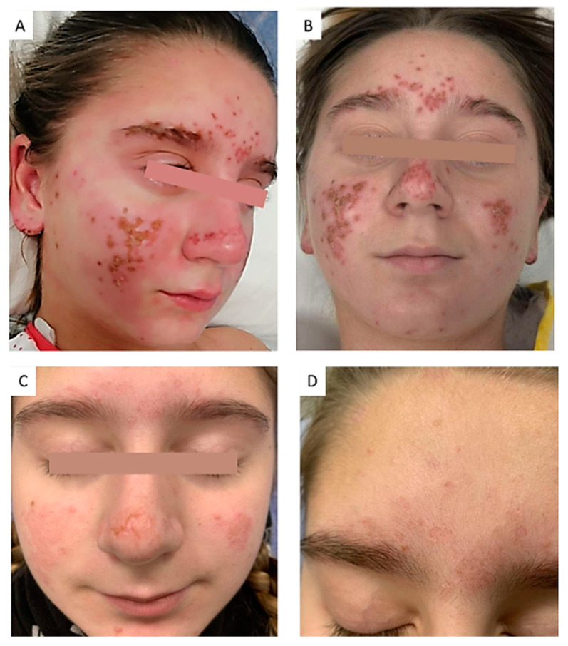
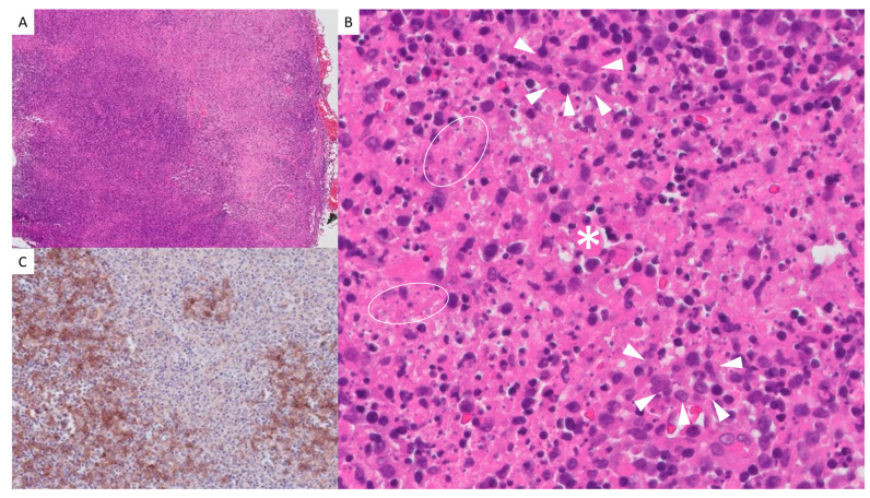
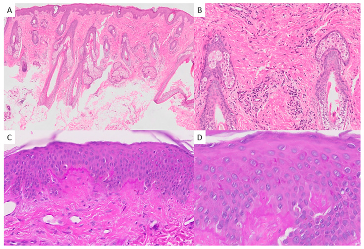
Kikuchi-Fujimoto disease (KFD) is a rare condition characterized by necrotizing lymphadenitis and fever, often associated with immune dysregulation. Histologically, it features necrotic foci with abundant histiocytes and plasmacytoid dendritic cells but notably lacks neutrophils and eosinophils. Recent evidence reveals a notable prevalence among pediatric patients, who may exhibit distinct features compared to adults. We reported the case of an 11-year-old girl presenting with persistent fever, cervical adenopathy, and a malar rash, leading to a diagnosis of KFD following lymph node biopsy, which revealed non-suppurative necrosis and histiocytic infiltration. Empirical treatment with antivirals and antibiotics was ineffective, but corticosteroid therapy achieved symptom remission. A literature review identified 48 relevant studies involving 386 pediatric cases, with histopathological findings consistent with classical descriptions of KFD. Cutaneous involvement was reported in 11.14% of cases, ranging from maculopapular rashes to lupus-like eruptions. Notable complications included the development of systemic lupus erythematous, Sjögren syndrome, and rare instances of hemophagocytic syndrome or central nervous system involvement. Kikuchi-Fujimoto disease should be considered in the differential diagnosis of pediatric patients presenting with fever and lymphadenopathy, taking into account a higher frequency of cutaneous manifestations in pediatric cases. A skin biopsy may be helpful in diagnosing KFD and provide valuable information regarding the potential risk of developing systemic lupus erythematosus in the future.

 求助内容:
求助内容: 应助结果提醒方式:


