Torben Fricke, Werner Kempf, Michael P Schön, Christina Mitteldorf
{"title":"淋巴瘤样丘疹病的组织学和免疫组织化学模式:对已发表病例的系统回顾。","authors":"Torben Fricke, Werner Kempf, Michael P Schön, Christina Mitteldorf","doi":"10.3390/dermatopathology12010006","DOIUrl":null,"url":null,"abstract":"<p><p>Based on histologic and genetic patterns, the current World Health Organization (WHO) classification distinguishes six subtypes of lymphomatoid papulosis (Lyp). The aim of our article was to analyze the frequency of histologic and immunohistochemical features of different Lyp subtypes reported in the literature. We used PubMed advanced search builder to systematically review and evaluate English and German literature of Lyp from 1968 to April 2022. We considered only papers in which histopathologic features were mentioned in detail. We identified 48 publications with a total of 518 cases. The diagnoses were based on the diagnostic criteria at the time of publication. In Lyp A and Lyp B a CD8+ phenotype was more often reported than expected (53% and 52%, respectively). A double positive phenotype (CD4+/CD8+) was found in 28% of Lyp E and a double negative (CD4-/CD8-) in 50% of Lyp with 6p25.3 rearrangement. High rates of folliculo- and syringotropism were reported in both Lyp A and B. Surprisingly, strong epidermotropism occurred in 20/38 (53%) cases reported as Lyp B and in 43/64 (67%) of Lyp D cases. The predominating phenotype in Lyp D was CD8+, while TIA-1/granzymeB/perforin expression was reported in 37/46 (80%), and CD56 was expressed in 13/47 (28%) of the investigated cases. The limitation of the data is due to the retrospective approach with diagnostic criteria changing over time and on a case selection in some publications. However, the data indicate that the Lyp subtypes overlap more than assumed. They also show that a prospective study is needed to obtain valid data on the frequency distribution of certain histopathologic criteria.</p>","PeriodicalId":42885,"journal":{"name":"Dermatopathology","volume":"12 1","pages":""},"PeriodicalIF":1.7000,"publicationDate":"2025-02-12","publicationTypes":"Journal Article","fieldsOfStudy":null,"isOpenAccess":false,"openAccessPdf":"https://www.ncbi.nlm.nih.gov/pmc/articles/PMC11861998/pdf/","citationCount":"0","resultStr":"{\"title\":\"Histologic and Immunohistochemical Patterns in Lymphomatoid Papulosis: A Systematic Review of Published Cases.\",\"authors\":\"Torben Fricke, Werner Kempf, Michael P Schön, Christina Mitteldorf\",\"doi\":\"10.3390/dermatopathology12010006\",\"DOIUrl\":null,\"url\":null,\"abstract\":\"<p><p>Based on histologic and genetic patterns, the current World Health Organization (WHO) classification distinguishes six subtypes of lymphomatoid papulosis (Lyp). The aim of our article was to analyze the frequency of histologic and immunohistochemical features of different Lyp subtypes reported in the literature. We used PubMed advanced search builder to systematically review and evaluate English and German literature of Lyp from 1968 to April 2022. We considered only papers in which histopathologic features were mentioned in detail. We identified 48 publications with a total of 518 cases. The diagnoses were based on the diagnostic criteria at the time of publication. In Lyp A and Lyp B a CD8+ phenotype was more often reported than expected (53% and 52%, respectively). A double positive phenotype (CD4+/CD8+) was found in 28% of Lyp E and a double negative (CD4-/CD8-) in 50% of Lyp with 6p25.3 rearrangement. High rates of folliculo- and syringotropism were reported in both Lyp A and B. Surprisingly, strong epidermotropism occurred in 20/38 (53%) cases reported as Lyp B and in 43/64 (67%) of Lyp D cases. The predominating phenotype in Lyp D was CD8+, while TIA-1/granzymeB/perforin expression was reported in 37/46 (80%), and CD56 was expressed in 13/47 (28%) of the investigated cases. The limitation of the data is due to the retrospective approach with diagnostic criteria changing over time and on a case selection in some publications. However, the data indicate that the Lyp subtypes overlap more than assumed. They also show that a prospective study is needed to obtain valid data on the frequency distribution of certain histopathologic criteria.</p>\",\"PeriodicalId\":42885,\"journal\":{\"name\":\"Dermatopathology\",\"volume\":\"12 1\",\"pages\":\"\"},\"PeriodicalIF\":1.7000,\"publicationDate\":\"2025-02-12\",\"publicationTypes\":\"Journal Article\",\"fieldsOfStudy\":null,\"isOpenAccess\":false,\"openAccessPdf\":\"https://www.ncbi.nlm.nih.gov/pmc/articles/PMC11861998/pdf/\",\"citationCount\":\"0\",\"resultStr\":null,\"platform\":\"Semanticscholar\",\"paperid\":null,\"PeriodicalName\":\"Dermatopathology\",\"FirstCategoryId\":\"1085\",\"ListUrlMain\":\"https://doi.org/10.3390/dermatopathology12010006\",\"RegionNum\":0,\"RegionCategory\":null,\"ArticlePicture\":[],\"TitleCN\":null,\"AbstractTextCN\":null,\"PMCID\":null,\"EPubDate\":\"\",\"PubModel\":\"\",\"JCR\":\"Q3\",\"JCRName\":\"DERMATOLOGY\",\"Score\":null,\"Total\":0}","platform":"Semanticscholar","paperid":null,"PeriodicalName":"Dermatopathology","FirstCategoryId":"1085","ListUrlMain":"https://doi.org/10.3390/dermatopathology12010006","RegionNum":0,"RegionCategory":null,"ArticlePicture":[],"TitleCN":null,"AbstractTextCN":null,"PMCID":null,"EPubDate":"","PubModel":"","JCR":"Q3","JCRName":"DERMATOLOGY","Score":null,"Total":0}
Histologic and Immunohistochemical Patterns in Lymphomatoid Papulosis: A Systematic Review of Published Cases.
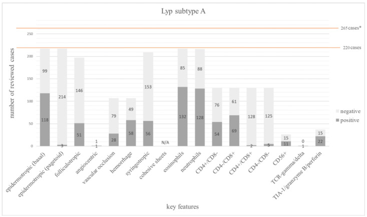
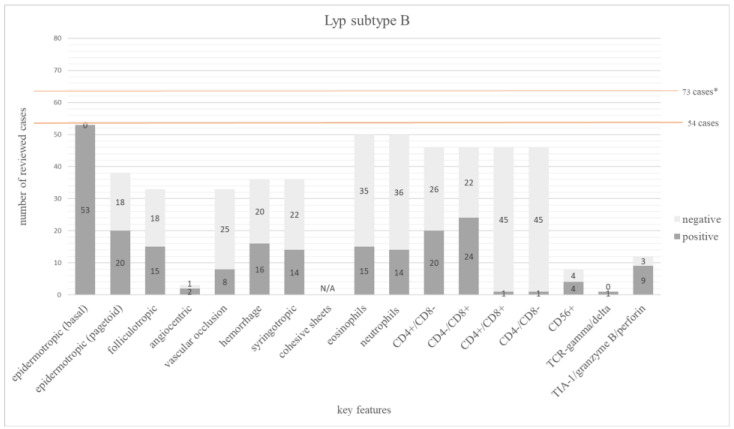
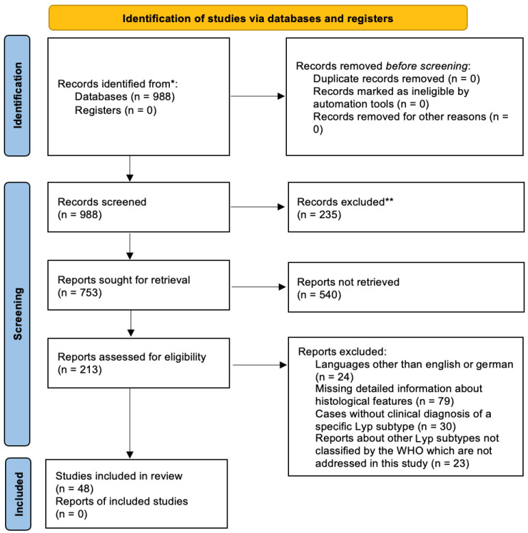
Based on histologic and genetic patterns, the current World Health Organization (WHO) classification distinguishes six subtypes of lymphomatoid papulosis (Lyp). The aim of our article was to analyze the frequency of histologic and immunohistochemical features of different Lyp subtypes reported in the literature. We used PubMed advanced search builder to systematically review and evaluate English and German literature of Lyp from 1968 to April 2022. We considered only papers in which histopathologic features were mentioned in detail. We identified 48 publications with a total of 518 cases. The diagnoses were based on the diagnostic criteria at the time of publication. In Lyp A and Lyp B a CD8+ phenotype was more often reported than expected (53% and 52%, respectively). A double positive phenotype (CD4+/CD8+) was found in 28% of Lyp E and a double negative (CD4-/CD8-) in 50% of Lyp with 6p25.3 rearrangement. High rates of folliculo- and syringotropism were reported in both Lyp A and B. Surprisingly, strong epidermotropism occurred in 20/38 (53%) cases reported as Lyp B and in 43/64 (67%) of Lyp D cases. The predominating phenotype in Lyp D was CD8+, while TIA-1/granzymeB/perforin expression was reported in 37/46 (80%), and CD56 was expressed in 13/47 (28%) of the investigated cases. The limitation of the data is due to the retrospective approach with diagnostic criteria changing over time and on a case selection in some publications. However, the data indicate that the Lyp subtypes overlap more than assumed. They also show that a prospective study is needed to obtain valid data on the frequency distribution of certain histopathologic criteria.

 求助内容:
求助内容: 应助结果提醒方式:


