Peng Yan, Zewen Wang, Yufeng Wang, Yongliang Liu, Anna Tong, Meili Sun
{"title":"胸腔放疗后同时服用抗 PD-1 与连续服用抗 PD-1 的比较分析:对肺组织损伤的影响","authors":"Peng Yan, Zewen Wang, Yufeng Wang, Yongliang Liu, Anna Tong, Meili Sun","doi":"10.1177/15593258251322324","DOIUrl":null,"url":null,"abstract":"<p><p>The combination of thoracic radiotherapy and immune checkpoint inhibitors (ICIs) had demonstrated a synergistic therapeutic effect, albeit with the occurrence of overlapping pulmonary toxicities. We established a mouse model using programmed cell death protein-1 (PD-1) antibody at different time points after thoracic radiotherapy. Hematoxylin and eosin (HE) staining, as well as TUNEL staining, were utilized for the morphological assessment of lung tissue damage. Inflammatory cells and cytokines present in bronchoalveolar lavage fluid (BALF) were analyzed using flow cytometry and cytometric bead array immunoassay (CBA). Additionally, immunohistochemistry (IHC) and immunofluorescence (IF) staining were conducted to observe the infiltration of inflammatory cells in lung tissue. Immediate administration of PD-1 antibody after thoracic radiotherapy resulted in more severe lung tissue injury compared to delayed administration. Concurrent treatment led to an increase in lymphocytes and neutrophils in BALF, as well as higher levels of inflammatory cytokines. IHC and IF analysis revealed that neutrophils, macrophages, and lymphocytes were more prominent in the concurrent treatment group. A more severe lung injury occurred when PD-1 antibody was given simultaneously with thoracic radiotherapy, possibly due to increased inflammation caused by the combination treatment.</p>","PeriodicalId":11285,"journal":{"name":"Dose-Response","volume":"23 1","pages":"15593258251322324"},"PeriodicalIF":2.4000,"publicationDate":"2025-02-20","publicationTypes":"Journal Article","fieldsOfStudy":null,"isOpenAccess":false,"openAccessPdf":"https://www.ncbi.nlm.nih.gov/pmc/articles/PMC11840853/pdf/","citationCount":"0","resultStr":"{\"title\":\"Comparative Analysis of Concurrent vs Sequential Administration of anti-PD-1 Following Thoracic Radiotherapy: Impact on Lung Tissue Damage.\",\"authors\":\"Peng Yan, Zewen Wang, Yufeng Wang, Yongliang Liu, Anna Tong, Meili Sun\",\"doi\":\"10.1177/15593258251322324\",\"DOIUrl\":null,\"url\":null,\"abstract\":\"<p><p>The combination of thoracic radiotherapy and immune checkpoint inhibitors (ICIs) had demonstrated a synergistic therapeutic effect, albeit with the occurrence of overlapping pulmonary toxicities. We established a mouse model using programmed cell death protein-1 (PD-1) antibody at different time points after thoracic radiotherapy. Hematoxylin and eosin (HE) staining, as well as TUNEL staining, were utilized for the morphological assessment of lung tissue damage. Inflammatory cells and cytokines present in bronchoalveolar lavage fluid (BALF) were analyzed using flow cytometry and cytometric bead array immunoassay (CBA). Additionally, immunohistochemistry (IHC) and immunofluorescence (IF) staining were conducted to observe the infiltration of inflammatory cells in lung tissue. Immediate administration of PD-1 antibody after thoracic radiotherapy resulted in more severe lung tissue injury compared to delayed administration. Concurrent treatment led to an increase in lymphocytes and neutrophils in BALF, as well as higher levels of inflammatory cytokines. IHC and IF analysis revealed that neutrophils, macrophages, and lymphocytes were more prominent in the concurrent treatment group. A more severe lung injury occurred when PD-1 antibody was given simultaneously with thoracic radiotherapy, possibly due to increased inflammation caused by the combination treatment.</p>\",\"PeriodicalId\":11285,\"journal\":{\"name\":\"Dose-Response\",\"volume\":\"23 1\",\"pages\":\"15593258251322324\"},\"PeriodicalIF\":2.4000,\"publicationDate\":\"2025-02-20\",\"publicationTypes\":\"Journal Article\",\"fieldsOfStudy\":null,\"isOpenAccess\":false,\"openAccessPdf\":\"https://www.ncbi.nlm.nih.gov/pmc/articles/PMC11840853/pdf/\",\"citationCount\":\"0\",\"resultStr\":null,\"platform\":\"Semanticscholar\",\"paperid\":null,\"PeriodicalName\":\"Dose-Response\",\"FirstCategoryId\":\"3\",\"ListUrlMain\":\"https://doi.org/10.1177/15593258251322324\",\"RegionNum\":4,\"RegionCategory\":\"医学\",\"ArticlePicture\":[],\"TitleCN\":null,\"AbstractTextCN\":null,\"PMCID\":null,\"EPubDate\":\"2025/1/1 0:00:00\",\"PubModel\":\"eCollection\",\"JCR\":\"Q3\",\"JCRName\":\"PHARMACOLOGY & PHARMACY\",\"Score\":null,\"Total\":0}","platform":"Semanticscholar","paperid":null,"PeriodicalName":"Dose-Response","FirstCategoryId":"3","ListUrlMain":"https://doi.org/10.1177/15593258251322324","RegionNum":4,"RegionCategory":"医学","ArticlePicture":[],"TitleCN":null,"AbstractTextCN":null,"PMCID":null,"EPubDate":"2025/1/1 0:00:00","PubModel":"eCollection","JCR":"Q3","JCRName":"PHARMACOLOGY & PHARMACY","Score":null,"Total":0}
Comparative Analysis of Concurrent vs Sequential Administration of anti-PD-1 Following Thoracic Radiotherapy: Impact on Lung Tissue Damage.
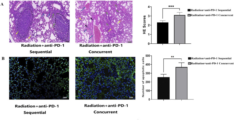
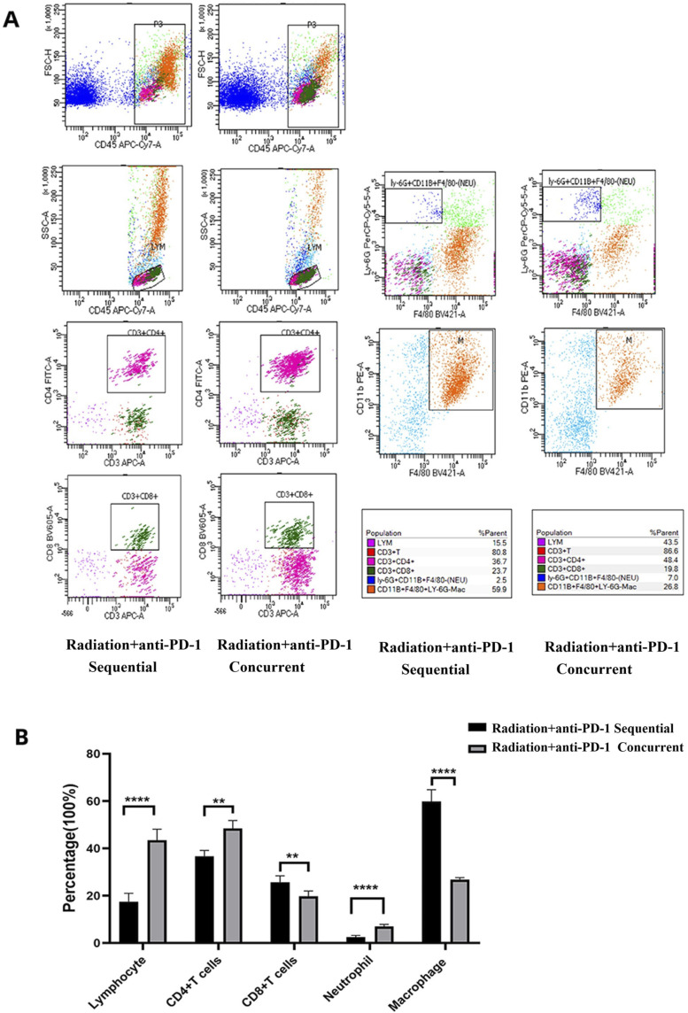
The combination of thoracic radiotherapy and immune checkpoint inhibitors (ICIs) had demonstrated a synergistic therapeutic effect, albeit with the occurrence of overlapping pulmonary toxicities. We established a mouse model using programmed cell death protein-1 (PD-1) antibody at different time points after thoracic radiotherapy. Hematoxylin and eosin (HE) staining, as well as TUNEL staining, were utilized for the morphological assessment of lung tissue damage. Inflammatory cells and cytokines present in bronchoalveolar lavage fluid (BALF) were analyzed using flow cytometry and cytometric bead array immunoassay (CBA). Additionally, immunohistochemistry (IHC) and immunofluorescence (IF) staining were conducted to observe the infiltration of inflammatory cells in lung tissue. Immediate administration of PD-1 antibody after thoracic radiotherapy resulted in more severe lung tissue injury compared to delayed administration. Concurrent treatment led to an increase in lymphocytes and neutrophils in BALF, as well as higher levels of inflammatory cytokines. IHC and IF analysis revealed that neutrophils, macrophages, and lymphocytes were more prominent in the concurrent treatment group. A more severe lung injury occurred when PD-1 antibody was given simultaneously with thoracic radiotherapy, possibly due to increased inflammation caused by the combination treatment.
Dose-ResponsePHARMACOLOGY & PHARMACY-RADIOLOGY, NUCLEAR MEDICINE & MEDICAL IMAGING
CiteScore
4.90
自引率
4.00%
发文量
140
审稿时长
>12 weeks
期刊介绍:
Dose-Response is an open access peer-reviewed online journal publishing original findings and commentaries on the occurrence of dose-response relationships across a broad range of disciplines. Particular interest focuses on experimental evidence providing mechanistic understanding of nonlinear dose-response relationships.

 求助内容:
求助内容: 应助结果提醒方式:


