Allison L. Horenberg, Yunke Ren, Eric Z. Zeng, Alexandra N. Rindone, Arvind P. Pathak, Warren L. Grayson
{"title":"三维成像显示小鼠颅骨神经血管结构随年龄的变化","authors":"Allison L. Horenberg, Yunke Ren, Eric Z. Zeng, Alexandra N. Rindone, Arvind P. Pathak, Warren L. Grayson","doi":"10.1038/s41413-025-00401-8","DOIUrl":null,"url":null,"abstract":"<p>Calvarial nerves, along with vasculature, influence skull formation during development and following injury, but it remains unclear how calvarial nerves are spatially distributed during postnatal growth and aging. Studying the spatial distribution of nerves in the skull remains a challenge due to a lack of methods to quantify 3D structures in intact bone. To visualize calvarial 3D neurovascular architecture, we imaged nerves and endothelial cells with lightsheet microscopy. We employed machine-learning-based segmentation to facilitate high-resolution characterization from post-natal day 0 (P0) to 80 weeks. We found that TUBB3<sup>+</sup> nerve density decreased with aging with the frontal bone demonstrating earlier onset age-related nerve loss than the parietal bone. In addition, nerves in the periosteum and dura mater exhibited similar yet distinct temporal patterns of nerve growth and loss. While no difference was observed in TUBB3<sup>+</sup> nerves during skeletal maturation (P0 → 12 weeks), we did observe an increase in the volume of unmyelinated nerves in the dura mater. Regarding calvarial vasculature, larger CD31<sup>hi</sup>Emcn<sup>-</sup> vessel fraction increased with aging, while CD31<sup>hi</sup>Emcn<sup>hi</sup> vessel fraction was reduced. Throughout all ages, calvarial nerves maintained a preferential spatial association with CD31<sup>hi</sup>Emcn<sup>hi</sup> vessels, however, this association decreased with aging. Additionally, we used a model of Apert syndrome to explore the impact of suture-related disease on neurovascular architecture. Collectively, this 3D, spatiotemporal characterization of calvarial nerves throughout the lifespan and provides new insights into age-induced neurovascular architecture.</p>","PeriodicalId":9134,"journal":{"name":"Bone Research","volume":"6 1","pages":""},"PeriodicalIF":15.0000,"publicationDate":"2025-02-21","publicationTypes":"Journal Article","fieldsOfStudy":null,"isOpenAccess":false,"openAccessPdf":"","citationCount":"0","resultStr":"{\"title\":\"3D imaging reveals changes in the neurovascular architecture of the murine calvarium with aging\",\"authors\":\"Allison L. Horenberg, Yunke Ren, Eric Z. Zeng, Alexandra N. Rindone, Arvind P. Pathak, Warren L. Grayson\",\"doi\":\"10.1038/s41413-025-00401-8\",\"DOIUrl\":null,\"url\":null,\"abstract\":\"<p>Calvarial nerves, along with vasculature, influence skull formation during development and following injury, but it remains unclear how calvarial nerves are spatially distributed during postnatal growth and aging. Studying the spatial distribution of nerves in the skull remains a challenge due to a lack of methods to quantify 3D structures in intact bone. To visualize calvarial 3D neurovascular architecture, we imaged nerves and endothelial cells with lightsheet microscopy. We employed machine-learning-based segmentation to facilitate high-resolution characterization from post-natal day 0 (P0) to 80 weeks. We found that TUBB3<sup>+</sup> nerve density decreased with aging with the frontal bone demonstrating earlier onset age-related nerve loss than the parietal bone. In addition, nerves in the periosteum and dura mater exhibited similar yet distinct temporal patterns of nerve growth and loss. While no difference was observed in TUBB3<sup>+</sup> nerves during skeletal maturation (P0 → 12 weeks), we did observe an increase in the volume of unmyelinated nerves in the dura mater. Regarding calvarial vasculature, larger CD31<sup>hi</sup>Emcn<sup>-</sup> vessel fraction increased with aging, while CD31<sup>hi</sup>Emcn<sup>hi</sup> vessel fraction was reduced. Throughout all ages, calvarial nerves maintained a preferential spatial association with CD31<sup>hi</sup>Emcn<sup>hi</sup> vessels, however, this association decreased with aging. Additionally, we used a model of Apert syndrome to explore the impact of suture-related disease on neurovascular architecture. Collectively, this 3D, spatiotemporal characterization of calvarial nerves throughout the lifespan and provides new insights into age-induced neurovascular architecture.</p>\",\"PeriodicalId\":9134,\"journal\":{\"name\":\"Bone Research\",\"volume\":\"6 1\",\"pages\":\"\"},\"PeriodicalIF\":15.0000,\"publicationDate\":\"2025-02-21\",\"publicationTypes\":\"Journal Article\",\"fieldsOfStudy\":null,\"isOpenAccess\":false,\"openAccessPdf\":\"\",\"citationCount\":\"0\",\"resultStr\":null,\"platform\":\"Semanticscholar\",\"paperid\":null,\"PeriodicalName\":\"Bone Research\",\"FirstCategoryId\":\"3\",\"ListUrlMain\":\"https://doi.org/10.1038/s41413-025-00401-8\",\"RegionNum\":1,\"RegionCategory\":\"医学\",\"ArticlePicture\":[],\"TitleCN\":null,\"AbstractTextCN\":null,\"PMCID\":null,\"EPubDate\":\"\",\"PubModel\":\"\",\"JCR\":\"Q1\",\"JCRName\":\"CELL & TISSUE ENGINEERING\",\"Score\":null,\"Total\":0}","platform":"Semanticscholar","paperid":null,"PeriodicalName":"Bone Research","FirstCategoryId":"3","ListUrlMain":"https://doi.org/10.1038/s41413-025-00401-8","RegionNum":1,"RegionCategory":"医学","ArticlePicture":[],"TitleCN":null,"AbstractTextCN":null,"PMCID":null,"EPubDate":"","PubModel":"","JCR":"Q1","JCRName":"CELL & TISSUE ENGINEERING","Score":null,"Total":0}
3D imaging reveals changes in the neurovascular architecture of the murine calvarium with aging
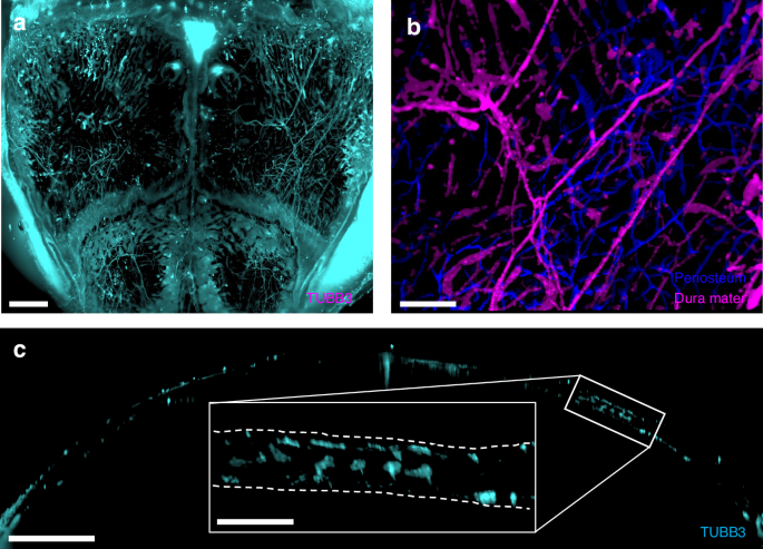
Calvarial nerves, along with vasculature, influence skull formation during development and following injury, but it remains unclear how calvarial nerves are spatially distributed during postnatal growth and aging. Studying the spatial distribution of nerves in the skull remains a challenge due to a lack of methods to quantify 3D structures in intact bone. To visualize calvarial 3D neurovascular architecture, we imaged nerves and endothelial cells with lightsheet microscopy. We employed machine-learning-based segmentation to facilitate high-resolution characterization from post-natal day 0 (P0) to 80 weeks. We found that TUBB3+ nerve density decreased with aging with the frontal bone demonstrating earlier onset age-related nerve loss than the parietal bone. In addition, nerves in the periosteum and dura mater exhibited similar yet distinct temporal patterns of nerve growth and loss. While no difference was observed in TUBB3+ nerves during skeletal maturation (P0 → 12 weeks), we did observe an increase in the volume of unmyelinated nerves in the dura mater. Regarding calvarial vasculature, larger CD31hiEmcn- vessel fraction increased with aging, while CD31hiEmcnhi vessel fraction was reduced. Throughout all ages, calvarial nerves maintained a preferential spatial association with CD31hiEmcnhi vessels, however, this association decreased with aging. Additionally, we used a model of Apert syndrome to explore the impact of suture-related disease on neurovascular architecture. Collectively, this 3D, spatiotemporal characterization of calvarial nerves throughout the lifespan and provides new insights into age-induced neurovascular architecture.
期刊介绍:
Established in 2013, Bone Research is a newly-founded English-language periodical that centers on the basic and clinical facets of bone biology, pathophysiology, and regeneration. It is dedicated to championing key findings emerging from both basic investigations and clinical research concerning bone-related topics. The journal's objective is to globally disseminate research in bone-related physiology, pathology, diseases, and treatment, contributing to the advancement of knowledge in this field.
 求助内容:
求助内容: 应助结果提醒方式:


