Giyeon Kim, Seonmi Kang, Junehee Seo, Kangmoon Seo
{"title":"睑缘厚度与睑腺功能障碍犬睑缘特征的关系。","authors":"Giyeon Kim, Seonmi Kang, Junehee Seo, Kangmoon Seo","doi":"10.1111/vop.13326","DOIUrl":null,"url":null,"abstract":"<p><strong>Objective: </strong>To measure the eyelid margin thickness (LMT) in dogs with meibomian gland dysfunction (MGD) and evaluate its correlation with meibomian gland (MG) morphology.</p><p><strong>Animals studied: </strong>Fifty-nine client-owned dogs.</p><p><strong>Procedure: </strong>The LMT was measured on slit lamp biomicroscopy images and divided into groups of 1 to 4, from the thinnest to thickest, based on quartiles. MG morphology, including distortion, thickening, shortening, and dropout, was evaluated using noninvasive infrared meibography. The LMT and meibography results were compared between the MGD and normal groups. Statistical analysis was performed to determine the correlation between LMT and MG morphology.</p><p><strong>Results: </strong>The mean LMT was significantly greater in the MGD group (1.18 ± 0.19 mm) than the normal group (1.00 ± 0.13 mm) and was positively correlated with MG loss (p < 0.01). The LMT was thicker in dogs over 12 years (1.25 ± 0.20 mm). The LMT group 4 (≥ 1.26 mm) had the highest percentage of abnormal MG (95.7%) and MG loss area (37%). The total abnormal MG ratio and thickened MG ratio were significantly higher in the LMT group 4. LMT ≥ 1.20 mm was identified as a potential indicator for MG loss area of more than one-third.</p><p><strong>Conclusions: </strong>Eyelids with thick LMT had more abnormal MG morphology, including thickening and dropout. An LMT ≥ 1.20 mm could be a criterion to suspect MGD. Thus, the LMT could be a simple screening tool to predict MG loss and might aid in the diagnosis and early management of MGD with a sensitivity of 0.645 and a specificity of 0.768.</p>","PeriodicalId":23836,"journal":{"name":"Veterinary ophthalmology","volume":" ","pages":"847-854"},"PeriodicalIF":1.3000,"publicationDate":"2025-09-01","publicationTypes":"Journal Article","fieldsOfStudy":null,"isOpenAccess":false,"openAccessPdf":"https://www.ncbi.nlm.nih.gov/pmc/articles/PMC12488535/pdf/","citationCount":"0","resultStr":"{\"title\":\"Association of Eyelid Margin Thickness and Meibography in Dogs With Meibomian Gland Dysfunction.\",\"authors\":\"Giyeon Kim, Seonmi Kang, Junehee Seo, Kangmoon Seo\",\"doi\":\"10.1111/vop.13326\",\"DOIUrl\":null,\"url\":null,\"abstract\":\"<p><strong>Objective: </strong>To measure the eyelid margin thickness (LMT) in dogs with meibomian gland dysfunction (MGD) and evaluate its correlation with meibomian gland (MG) morphology.</p><p><strong>Animals studied: </strong>Fifty-nine client-owned dogs.</p><p><strong>Procedure: </strong>The LMT was measured on slit lamp biomicroscopy images and divided into groups of 1 to 4, from the thinnest to thickest, based on quartiles. MG morphology, including distortion, thickening, shortening, and dropout, was evaluated using noninvasive infrared meibography. The LMT and meibography results were compared between the MGD and normal groups. Statistical analysis was performed to determine the correlation between LMT and MG morphology.</p><p><strong>Results: </strong>The mean LMT was significantly greater in the MGD group (1.18 ± 0.19 mm) than the normal group (1.00 ± 0.13 mm) and was positively correlated with MG loss (p < 0.01). The LMT was thicker in dogs over 12 years (1.25 ± 0.20 mm). The LMT group 4 (≥ 1.26 mm) had the highest percentage of abnormal MG (95.7%) and MG loss area (37%). The total abnormal MG ratio and thickened MG ratio were significantly higher in the LMT group 4. LMT ≥ 1.20 mm was identified as a potential indicator for MG loss area of more than one-third.</p><p><strong>Conclusions: </strong>Eyelids with thick LMT had more abnormal MG morphology, including thickening and dropout. An LMT ≥ 1.20 mm could be a criterion to suspect MGD. Thus, the LMT could be a simple screening tool to predict MG loss and might aid in the diagnosis and early management of MGD with a sensitivity of 0.645 and a specificity of 0.768.</p>\",\"PeriodicalId\":23836,\"journal\":{\"name\":\"Veterinary ophthalmology\",\"volume\":\" \",\"pages\":\"847-854\"},\"PeriodicalIF\":1.3000,\"publicationDate\":\"2025-09-01\",\"publicationTypes\":\"Journal Article\",\"fieldsOfStudy\":null,\"isOpenAccess\":false,\"openAccessPdf\":\"https://www.ncbi.nlm.nih.gov/pmc/articles/PMC12488535/pdf/\",\"citationCount\":\"0\",\"resultStr\":null,\"platform\":\"Semanticscholar\",\"paperid\":null,\"PeriodicalName\":\"Veterinary ophthalmology\",\"FirstCategoryId\":\"97\",\"ListUrlMain\":\"https://doi.org/10.1111/vop.13326\",\"RegionNum\":4,\"RegionCategory\":\"农林科学\",\"ArticlePicture\":[],\"TitleCN\":null,\"AbstractTextCN\":null,\"PMCID\":null,\"EPubDate\":\"2025/2/18 0:00:00\",\"PubModel\":\"Epub\",\"JCR\":\"Q2\",\"JCRName\":\"VETERINARY SCIENCES\",\"Score\":null,\"Total\":0}","platform":"Semanticscholar","paperid":null,"PeriodicalName":"Veterinary ophthalmology","FirstCategoryId":"97","ListUrlMain":"https://doi.org/10.1111/vop.13326","RegionNum":4,"RegionCategory":"农林科学","ArticlePicture":[],"TitleCN":null,"AbstractTextCN":null,"PMCID":null,"EPubDate":"2025/2/18 0:00:00","PubModel":"Epub","JCR":"Q2","JCRName":"VETERINARY SCIENCES","Score":null,"Total":0}
Association of Eyelid Margin Thickness and Meibography in Dogs With Meibomian Gland Dysfunction.
Objective: To measure the eyelid margin thickness (LMT) in dogs with meibomian gland dysfunction (MGD) and evaluate its correlation with meibomian gland (MG) morphology.
Animals studied: Fifty-nine client-owned dogs.
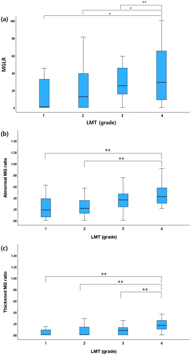
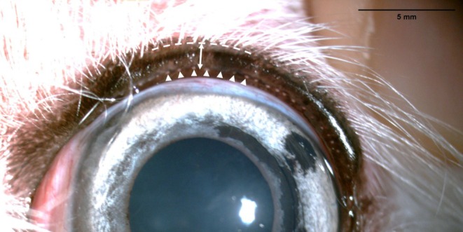
Procedure: The LMT was measured on slit lamp biomicroscopy images and divided into groups of 1 to 4, from the thinnest to thickest, based on quartiles. MG morphology, including distortion, thickening, shortening, and dropout, was evaluated using noninvasive infrared meibography. The LMT and meibography results were compared between the MGD and normal groups. Statistical analysis was performed to determine the correlation between LMT and MG morphology.
Results: The mean LMT was significantly greater in the MGD group (1.18 ± 0.19 mm) than the normal group (1.00 ± 0.13 mm) and was positively correlated with MG loss (p < 0.01). The LMT was thicker in dogs over 12 years (1.25 ± 0.20 mm). The LMT group 4 (≥ 1.26 mm) had the highest percentage of abnormal MG (95.7%) and MG loss area (37%). The total abnormal MG ratio and thickened MG ratio were significantly higher in the LMT group 4. LMT ≥ 1.20 mm was identified as a potential indicator for MG loss area of more than one-third.
Conclusions: Eyelids with thick LMT had more abnormal MG morphology, including thickening and dropout. An LMT ≥ 1.20 mm could be a criterion to suspect MGD. Thus, the LMT could be a simple screening tool to predict MG loss and might aid in the diagnosis and early management of MGD with a sensitivity of 0.645 and a specificity of 0.768.
期刊介绍:
Veterinary Ophthalmology is a peer-reviewed, international journal that welcomes submission of manuscripts directed towards academic researchers of veterinary ophthalmology, specialists and general practitioners with a strong ophthalmology interest. Articles include those relating to all aspects of:
Clinical and investigational veterinary and comparative ophthalmology;
Prospective and retrospective studies or reviews of naturally occurring ocular disease in veterinary species;
Experimental models of both animal and human ocular disease in veterinary species;
Anatomic studies of the animal eye;
Physiological studies of the animal eye;
Pharmacological studies of the animal eye.


 求助内容:
求助内容: 应助结果提醒方式:


