Eva Blondeel, Arne Peirsman, Stephanie Vermeulen, Filippo Piccinini, Felix De Vuyst, Diogo Estêvão, Sayida Al-Jamei, Martina Bedeschi, Gastone Castellani, Tânia Cruz, Sándor Dedeyne, Maria José Oliveira, Satoru Kawakita, Huu Tuan Nguyen, Leoni A Kunz-Schughart, Soojung Lee, Noemi Marino, Patrick Steigemann, Shuichi Takayama, Anna Tesei, Nina Zablowsky, Phillip Blondeel, Olivier De Wever
{"title":"用于三维细胞培养形态计量学分析的球形光显微镜图像图谱。","authors":"Eva Blondeel, Arne Peirsman, Stephanie Vermeulen, Filippo Piccinini, Felix De Vuyst, Diogo Estêvão, Sayida Al-Jamei, Martina Bedeschi, Gastone Castellani, Tânia Cruz, Sándor Dedeyne, Maria José Oliveira, Satoru Kawakita, Huu Tuan Nguyen, Leoni A Kunz-Schughart, Soojung Lee, Noemi Marino, Patrick Steigemann, Shuichi Takayama, Anna Tesei, Nina Zablowsky, Phillip Blondeel, Olivier De Wever","doi":"10.1038/s41597-025-04441-x","DOIUrl":null,"url":null,"abstract":"<p><p>The application of three-dimensional (3D) cell cultures such as spheroids and organoids is growing in popularity both in academia and industry. However, morphology of the 3D architecture remains remarkably understudied. Here, we introduce an open-access Spheroid Light Microscopy Image Atlas (SLiMIA) that can serve as a training set for morphology studies of 3D cell cultures. We provide images with a variety of metadata: 9 microscopes, 47 cell lines, 8 culture media, 4 spheroid formation methods and multiple cell seeding densities; totalling approximately 8,000 images of spheroids. This comprehensive dataset can guide spheroid researchers and promote economizing of resources by advancing 3D cell culture optimization, standardization and implementation by the community at large. Considering the exponentially growing interest in spheroid morphometrical analyses and the emerging technological possibilities to do so, this atlas can be applied to train and develop image segmentation models to deepen our understanding of 3D spheroid morphometry in biomedical research.</p>","PeriodicalId":21597,"journal":{"name":"Scientific Data","volume":"12 1","pages":"283"},"PeriodicalIF":6.9000,"publicationDate":"2025-02-17","publicationTypes":"Journal Article","fieldsOfStudy":null,"isOpenAccess":false,"openAccessPdf":"https://www.ncbi.nlm.nih.gov/pmc/articles/PMC11833042/pdf/","citationCount":"0","resultStr":"{\"title\":\"The Spheroid Light Microscopy Image Atlas for morphometrical analysis of three-dimensional cell cultures.\",\"authors\":\"Eva Blondeel, Arne Peirsman, Stephanie Vermeulen, Filippo Piccinini, Felix De Vuyst, Diogo Estêvão, Sayida Al-Jamei, Martina Bedeschi, Gastone Castellani, Tânia Cruz, Sándor Dedeyne, Maria José Oliveira, Satoru Kawakita, Huu Tuan Nguyen, Leoni A Kunz-Schughart, Soojung Lee, Noemi Marino, Patrick Steigemann, Shuichi Takayama, Anna Tesei, Nina Zablowsky, Phillip Blondeel, Olivier De Wever\",\"doi\":\"10.1038/s41597-025-04441-x\",\"DOIUrl\":null,\"url\":null,\"abstract\":\"<p><p>The application of three-dimensional (3D) cell cultures such as spheroids and organoids is growing in popularity both in academia and industry. However, morphology of the 3D architecture remains remarkably understudied. Here, we introduce an open-access Spheroid Light Microscopy Image Atlas (SLiMIA) that can serve as a training set for morphology studies of 3D cell cultures. We provide images with a variety of metadata: 9 microscopes, 47 cell lines, 8 culture media, 4 spheroid formation methods and multiple cell seeding densities; totalling approximately 8,000 images of spheroids. This comprehensive dataset can guide spheroid researchers and promote economizing of resources by advancing 3D cell culture optimization, standardization and implementation by the community at large. Considering the exponentially growing interest in spheroid morphometrical analyses and the emerging technological possibilities to do so, this atlas can be applied to train and develop image segmentation models to deepen our understanding of 3D spheroid morphometry in biomedical research.</p>\",\"PeriodicalId\":21597,\"journal\":{\"name\":\"Scientific Data\",\"volume\":\"12 1\",\"pages\":\"283\"},\"PeriodicalIF\":6.9000,\"publicationDate\":\"2025-02-17\",\"publicationTypes\":\"Journal Article\",\"fieldsOfStudy\":null,\"isOpenAccess\":false,\"openAccessPdf\":\"https://www.ncbi.nlm.nih.gov/pmc/articles/PMC11833042/pdf/\",\"citationCount\":\"0\",\"resultStr\":null,\"platform\":\"Semanticscholar\",\"paperid\":null,\"PeriodicalName\":\"Scientific Data\",\"FirstCategoryId\":\"103\",\"ListUrlMain\":\"https://doi.org/10.1038/s41597-025-04441-x\",\"RegionNum\":2,\"RegionCategory\":\"综合性期刊\",\"ArticlePicture\":[],\"TitleCN\":null,\"AbstractTextCN\":null,\"PMCID\":null,\"EPubDate\":\"\",\"PubModel\":\"\",\"JCR\":\"Q1\",\"JCRName\":\"MULTIDISCIPLINARY SCIENCES\",\"Score\":null,\"Total\":0}","platform":"Semanticscholar","paperid":null,"PeriodicalName":"Scientific Data","FirstCategoryId":"103","ListUrlMain":"https://doi.org/10.1038/s41597-025-04441-x","RegionNum":2,"RegionCategory":"综合性期刊","ArticlePicture":[],"TitleCN":null,"AbstractTextCN":null,"PMCID":null,"EPubDate":"","PubModel":"","JCR":"Q1","JCRName":"MULTIDISCIPLINARY SCIENCES","Score":null,"Total":0}
The Spheroid Light Microscopy Image Atlas for morphometrical analysis of three-dimensional cell cultures.
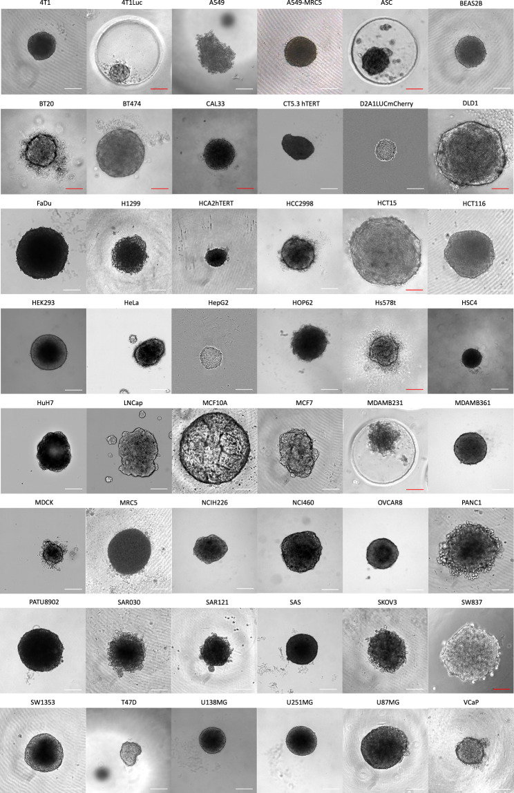
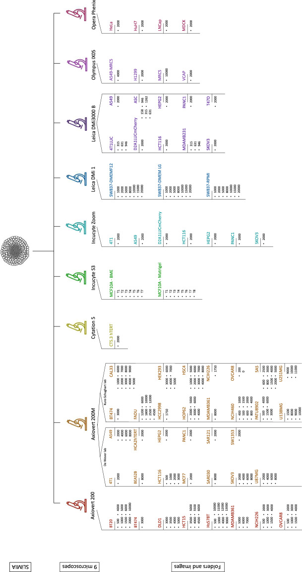
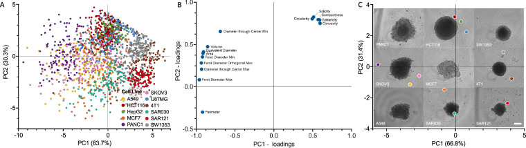
The application of three-dimensional (3D) cell cultures such as spheroids and organoids is growing in popularity both in academia and industry. However, morphology of the 3D architecture remains remarkably understudied. Here, we introduce an open-access Spheroid Light Microscopy Image Atlas (SLiMIA) that can serve as a training set for morphology studies of 3D cell cultures. We provide images with a variety of metadata: 9 microscopes, 47 cell lines, 8 culture media, 4 spheroid formation methods and multiple cell seeding densities; totalling approximately 8,000 images of spheroids. This comprehensive dataset can guide spheroid researchers and promote economizing of resources by advancing 3D cell culture optimization, standardization and implementation by the community at large. Considering the exponentially growing interest in spheroid morphometrical analyses and the emerging technological possibilities to do so, this atlas can be applied to train and develop image segmentation models to deepen our understanding of 3D spheroid morphometry in biomedical research.
期刊介绍:
Scientific Data is an open-access journal focused on data, publishing descriptions of research datasets and articles on data sharing across natural sciences, medicine, engineering, and social sciences. Its goal is to enhance the sharing and reuse of scientific data, encourage broader data sharing, and acknowledge those who share their data.
The journal primarily publishes Data Descriptors, which offer detailed descriptions of research datasets, including data collection methods and technical analyses validating data quality. These descriptors aim to facilitate data reuse rather than testing hypotheses or presenting new interpretations, methods, or in-depth analyses.
 求助内容:
求助内容: 应助结果提醒方式:


