Huda Al Ghadeer, Catherine Wang, Hind Alkatan, Rajiv Khandekar, Azza Al Maktabi, Deepak P Edward
{"title":"结膜淋巴血管扩张可能是淋巴静脉病变:临床病理和免疫组织化学研究。","authors":"Huda Al Ghadeer, Catherine Wang, Hind Alkatan, Rajiv Khandekar, Azza Al Maktabi, Deepak P Edward","doi":"10.4103/meajo.meajo_95_24","DOIUrl":null,"url":null,"abstract":"<p><strong>Purpose: </strong>To report the clinical presentation, histopathological and immunohistological features of conjunctival lymphatic-venous lesions.</p><p><strong>Methods: </strong>This was a retrospective review of 15 patients with symptomatic conjunctival lymphatic-venous lesions. The clinical features and histopathologic features of the excised tissues were reviewed. Immunohistochemical staining with antibodies against CD34 to label vascular endothelium and D2-40 to label lymphatic endothelium was performed.</p><p><strong>Results: </strong>All patients had unilateral conjunctival involvement. The mean age was 48.9 ± 18.9 years, with more women affected (67%). No patient had impaired visual acuity secondary to the lesion. The location of the lesion was temporal in 67% of patients. The presenting symptoms included conjunctival swelling, discomfort and/or foreign-body sensation, and tearing. All lesions were excised, and none recurred. All excised lesions showed dilated channels that were lined by a flattened endothelium staining positive with CD34, surrounded by edematous lamina propria. Larger ectatic lymphatic channels demonstrated scattered D2-40 staining in the endothelial cell lining and patchy CD34 staining within the endothelial cell cytoplasm. D2-40 and CD-34 immunoreactivity did not overlap in the same cells.</p><p><strong>Conclusion: </strong>The clinical features and outcomes of the lesions in this large cohort were similar to those reported in the literature. However, the mixed immunoreactivity of the endothelial cells lining these ectatic lymphatic channels in the conjunctiva suggests that these channels are lymphatic-venous lesions. We suggest that these channels be termed conjunctival lymphaticovenous malformation rather than lymphangiectasia, which suggests ectasia of existing lymphatics. Future studies are needed to understand these lesions and their histopathologic origins.</p>","PeriodicalId":18740,"journal":{"name":"Middle East African Journal of Ophthalmology","volume":"30 4","pages":"214-219"},"PeriodicalIF":0.3000,"publicationDate":"2024-12-02","publicationTypes":"Journal Article","fieldsOfStudy":null,"isOpenAccess":false,"openAccessPdf":"https://www.ncbi.nlm.nih.gov/pmc/articles/PMC11823533/pdf/","citationCount":"0","resultStr":"{\"title\":\"Conjunctival Lymphangiectasia is Likely Lymphatic-venous Lesions: A Clinicopathologic and Immunohistochemical Study.\",\"authors\":\"Huda Al Ghadeer, Catherine Wang, Hind Alkatan, Rajiv Khandekar, Azza Al Maktabi, Deepak P Edward\",\"doi\":\"10.4103/meajo.meajo_95_24\",\"DOIUrl\":null,\"url\":null,\"abstract\":\"<p><strong>Purpose: </strong>To report the clinical presentation, histopathological and immunohistological features of conjunctival lymphatic-venous lesions.</p><p><strong>Methods: </strong>This was a retrospective review of 15 patients with symptomatic conjunctival lymphatic-venous lesions. The clinical features and histopathologic features of the excised tissues were reviewed. Immunohistochemical staining with antibodies against CD34 to label vascular endothelium and D2-40 to label lymphatic endothelium was performed.</p><p><strong>Results: </strong>All patients had unilateral conjunctival involvement. The mean age was 48.9 ± 18.9 years, with more women affected (67%). No patient had impaired visual acuity secondary to the lesion. The location of the lesion was temporal in 67% of patients. The presenting symptoms included conjunctival swelling, discomfort and/or foreign-body sensation, and tearing. All lesions were excised, and none recurred. All excised lesions showed dilated channels that were lined by a flattened endothelium staining positive with CD34, surrounded by edematous lamina propria. Larger ectatic lymphatic channels demonstrated scattered D2-40 staining in the endothelial cell lining and patchy CD34 staining within the endothelial cell cytoplasm. D2-40 and CD-34 immunoreactivity did not overlap in the same cells.</p><p><strong>Conclusion: </strong>The clinical features and outcomes of the lesions in this large cohort were similar to those reported in the literature. However, the mixed immunoreactivity of the endothelial cells lining these ectatic lymphatic channels in the conjunctiva suggests that these channels are lymphatic-venous lesions. We suggest that these channels be termed conjunctival lymphaticovenous malformation rather than lymphangiectasia, which suggests ectasia of existing lymphatics. Future studies are needed to understand these lesions and their histopathologic origins.</p>\",\"PeriodicalId\":18740,\"journal\":{\"name\":\"Middle East African Journal of Ophthalmology\",\"volume\":\"30 4\",\"pages\":\"214-219\"},\"PeriodicalIF\":0.3000,\"publicationDate\":\"2024-12-02\",\"publicationTypes\":\"Journal Article\",\"fieldsOfStudy\":null,\"isOpenAccess\":false,\"openAccessPdf\":\"https://www.ncbi.nlm.nih.gov/pmc/articles/PMC11823533/pdf/\",\"citationCount\":\"0\",\"resultStr\":null,\"platform\":\"Semanticscholar\",\"paperid\":null,\"PeriodicalName\":\"Middle East African Journal of Ophthalmology\",\"FirstCategoryId\":\"1085\",\"ListUrlMain\":\"https://doi.org/10.4103/meajo.meajo_95_24\",\"RegionNum\":0,\"RegionCategory\":null,\"ArticlePicture\":[],\"TitleCN\":null,\"AbstractTextCN\":null,\"PMCID\":null,\"EPubDate\":\"2023/10/1 0:00:00\",\"PubModel\":\"eCollection\",\"JCR\":\"Q4\",\"JCRName\":\"OPHTHALMOLOGY\",\"Score\":null,\"Total\":0}","platform":"Semanticscholar","paperid":null,"PeriodicalName":"Middle East African Journal of Ophthalmology","FirstCategoryId":"1085","ListUrlMain":"https://doi.org/10.4103/meajo.meajo_95_24","RegionNum":0,"RegionCategory":null,"ArticlePicture":[],"TitleCN":null,"AbstractTextCN":null,"PMCID":null,"EPubDate":"2023/10/1 0:00:00","PubModel":"eCollection","JCR":"Q4","JCRName":"OPHTHALMOLOGY","Score":null,"Total":0}
Conjunctival Lymphangiectasia is Likely Lymphatic-venous Lesions: A Clinicopathologic and Immunohistochemical Study.
Purpose: To report the clinical presentation, histopathological and immunohistological features of conjunctival lymphatic-venous lesions.
Methods: This was a retrospective review of 15 patients with symptomatic conjunctival lymphatic-venous lesions. The clinical features and histopathologic features of the excised tissues were reviewed. Immunohistochemical staining with antibodies against CD34 to label vascular endothelium and D2-40 to label lymphatic endothelium was performed.
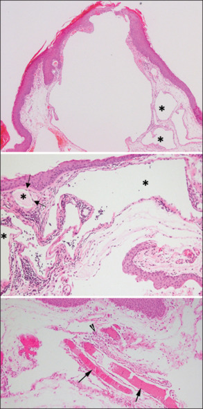
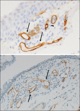
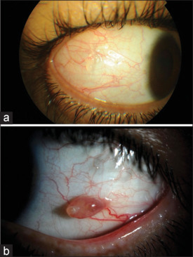
Results: All patients had unilateral conjunctival involvement. The mean age was 48.9 ± 18.9 years, with more women affected (67%). No patient had impaired visual acuity secondary to the lesion. The location of the lesion was temporal in 67% of patients. The presenting symptoms included conjunctival swelling, discomfort and/or foreign-body sensation, and tearing. All lesions were excised, and none recurred. All excised lesions showed dilated channels that were lined by a flattened endothelium staining positive with CD34, surrounded by edematous lamina propria. Larger ectatic lymphatic channels demonstrated scattered D2-40 staining in the endothelial cell lining and patchy CD34 staining within the endothelial cell cytoplasm. D2-40 and CD-34 immunoreactivity did not overlap in the same cells.
Conclusion: The clinical features and outcomes of the lesions in this large cohort were similar to those reported in the literature. However, the mixed immunoreactivity of the endothelial cells lining these ectatic lymphatic channels in the conjunctiva suggests that these channels are lymphatic-venous lesions. We suggest that these channels be termed conjunctival lymphaticovenous malformation rather than lymphangiectasia, which suggests ectasia of existing lymphatics. Future studies are needed to understand these lesions and their histopathologic origins.
期刊介绍:
The Middle East African Journal of Ophthalmology (MEAJO), published four times per year in print and online, is an official journal of the Middle East African Council of Ophthalmology (MEACO). It is an international, peer-reviewed journal whose mission includes publication of original research of interest to ophthalmologists in the Middle East and Africa, and to provide readers with high quality educational review articles from world-renown experts. MEAJO, previously known as Middle East Journal of Ophthalmology (MEJO) was founded by Dr Akef El Maghraby in 1993.

 求助内容:
求助内容: 应助结果提醒方式:


