E-O Sousa, N-A Mirsky, M Parra, V-V Nayak, B-L Silva, E-A Bonfante, N Tovar, P-G Coelho, L Witek
{"title":"利用高分子生物可吸收膜保存眼窝:临床前模型。","authors":"E-O Sousa, N-A Mirsky, M Parra, V-V Nayak, B-L Silva, E-A Bonfante, N Tovar, P-G Coelho, L Witek","doi":"10.4317/medoral.26938","DOIUrl":null,"url":null,"abstract":"<p><strong>Background: </strong>The preservation of the alveolar ridge following tooth extraction is crucial to prevent atrophy and maintain structural integrity, facilitating future dental rehabilitations. This study compared the use of two different polymeric, resorbable membranes: polylactic acid (PLA), and 5% polylactic acid + 95% polycaprolactone (PLA/PCL), relative to unassisted socket healing (negative control).</p><p><strong>Material and methods: </strong>A preclinical model involving healthy, skeletally mature beagles (n=7) were used in this study. Surface topography and thermal degradation of the membranes were assessed, followed by in vivo evaluation of socket preservation in extracted maxillary premolars. Histomorphometric analysis was employed to measure bone formation and total socket area. Data was analyzed through linear mixed models with fixed factor of treatment following a post-hoc comparison by the Tukey test. Ranked data of residual membrane presence and inflammatory infiltrate were analyzed through Kruskal-Wallis non-parametric test. All analyses were conducted with statistical significance set at p-value ≤ 0.05.</p><p><strong>Results: </strong>Surface topography depicted a distinctive fibrous network structure for PLA membrane relative to PLA/PCL which exhibited a more porous architecture. Thermal degradation behavior/profile, observed through TGA and DSC, for both membranes was similar. Histomorphometric analysis of bone formation within the induced socket yielded 36.1 ±7.7%, 35.6 ±7.2% and 32.8 ±7.7% for control, PLA and PLA/PCL groups, respectively, with no statistically significant differences between groups (p = 0.796). Analysis of total socket area (mean ± 95% confidence intervals) yielded significantly higher values for experimental groups, PLA (8.95 ± 1.7 mm2) and PLA/PCL (8.8 ± 1.76 mm2), relative to control (6.7 ± 1.8 mm2) (p = 0.041). Residual membrane, along with mild inflammatory infiltrate was observed after the healing period irrespective of membrane type utilized.</p><p><strong>Conclusions: </strong>Guided bone regeneration (GBR) with PLA and PLA/PCL membranes did not yield higher bone formation within the socket relative to the control group. However, an improvement in the preservation of the socket's architecture was observed.</p>","PeriodicalId":49016,"journal":{"name":"Medicina Oral Patologia Oral Y Cirugia Bucal","volume":" ","pages":"e288-e296"},"PeriodicalIF":2.1000,"publicationDate":"2025-03-01","publicationTypes":"Journal Article","fieldsOfStudy":null,"isOpenAccess":false,"openAccessPdf":"https://www.ncbi.nlm.nih.gov/pmc/articles/PMC11972654/pdf/","citationCount":"0","resultStr":"{\"title\":\"Socket preservation utilizing polymeric bioresorbable membranes: a preclinical model.\",\"authors\":\"E-O Sousa, N-A Mirsky, M Parra, V-V Nayak, B-L Silva, E-A Bonfante, N Tovar, P-G Coelho, L Witek\",\"doi\":\"10.4317/medoral.26938\",\"DOIUrl\":null,\"url\":null,\"abstract\":\"<p><strong>Background: </strong>The preservation of the alveolar ridge following tooth extraction is crucial to prevent atrophy and maintain structural integrity, facilitating future dental rehabilitations. This study compared the use of two different polymeric, resorbable membranes: polylactic acid (PLA), and 5% polylactic acid + 95% polycaprolactone (PLA/PCL), relative to unassisted socket healing (negative control).</p><p><strong>Material and methods: </strong>A preclinical model involving healthy, skeletally mature beagles (n=7) were used in this study. Surface topography and thermal degradation of the membranes were assessed, followed by in vivo evaluation of socket preservation in extracted maxillary premolars. Histomorphometric analysis was employed to measure bone formation and total socket area. Data was analyzed through linear mixed models with fixed factor of treatment following a post-hoc comparison by the Tukey test. Ranked data of residual membrane presence and inflammatory infiltrate were analyzed through Kruskal-Wallis non-parametric test. All analyses were conducted with statistical significance set at p-value ≤ 0.05.</p><p><strong>Results: </strong>Surface topography depicted a distinctive fibrous network structure for PLA membrane relative to PLA/PCL which exhibited a more porous architecture. Thermal degradation behavior/profile, observed through TGA and DSC, for both membranes was similar. Histomorphometric analysis of bone formation within the induced socket yielded 36.1 ±7.7%, 35.6 ±7.2% and 32.8 ±7.7% for control, PLA and PLA/PCL groups, respectively, with no statistically significant differences between groups (p = 0.796). Analysis of total socket area (mean ± 95% confidence intervals) yielded significantly higher values for experimental groups, PLA (8.95 ± 1.7 mm2) and PLA/PCL (8.8 ± 1.76 mm2), relative to control (6.7 ± 1.8 mm2) (p = 0.041). Residual membrane, along with mild inflammatory infiltrate was observed after the healing period irrespective of membrane type utilized.</p><p><strong>Conclusions: </strong>Guided bone regeneration (GBR) with PLA and PLA/PCL membranes did not yield higher bone formation within the socket relative to the control group. However, an improvement in the preservation of the socket's architecture was observed.</p>\",\"PeriodicalId\":49016,\"journal\":{\"name\":\"Medicina Oral Patologia Oral Y Cirugia Bucal\",\"volume\":\" \",\"pages\":\"e288-e296\"},\"PeriodicalIF\":2.1000,\"publicationDate\":\"2025-03-01\",\"publicationTypes\":\"Journal Article\",\"fieldsOfStudy\":null,\"isOpenAccess\":false,\"openAccessPdf\":\"https://www.ncbi.nlm.nih.gov/pmc/articles/PMC11972654/pdf/\",\"citationCount\":\"0\",\"resultStr\":null,\"platform\":\"Semanticscholar\",\"paperid\":null,\"PeriodicalName\":\"Medicina Oral Patologia Oral Y Cirugia Bucal\",\"FirstCategoryId\":\"3\",\"ListUrlMain\":\"https://doi.org/10.4317/medoral.26938\",\"RegionNum\":3,\"RegionCategory\":\"医学\",\"ArticlePicture\":[],\"TitleCN\":null,\"AbstractTextCN\":null,\"PMCID\":null,\"EPubDate\":\"\",\"PubModel\":\"\",\"JCR\":\"Q2\",\"JCRName\":\"DENTISTRY, ORAL SURGERY & MEDICINE\",\"Score\":null,\"Total\":0}","platform":"Semanticscholar","paperid":null,"PeriodicalName":"Medicina Oral Patologia Oral Y Cirugia Bucal","FirstCategoryId":"3","ListUrlMain":"https://doi.org/10.4317/medoral.26938","RegionNum":3,"RegionCategory":"医学","ArticlePicture":[],"TitleCN":null,"AbstractTextCN":null,"PMCID":null,"EPubDate":"","PubModel":"","JCR":"Q2","JCRName":"DENTISTRY, ORAL SURGERY & MEDICINE","Score":null,"Total":0}
Socket preservation utilizing polymeric bioresorbable membranes: a preclinical model.
Background: The preservation of the alveolar ridge following tooth extraction is crucial to prevent atrophy and maintain structural integrity, facilitating future dental rehabilitations. This study compared the use of two different polymeric, resorbable membranes: polylactic acid (PLA), and 5% polylactic acid + 95% polycaprolactone (PLA/PCL), relative to unassisted socket healing (negative control).
Material and methods: A preclinical model involving healthy, skeletally mature beagles (n=7) were used in this study. Surface topography and thermal degradation of the membranes were assessed, followed by in vivo evaluation of socket preservation in extracted maxillary premolars. Histomorphometric analysis was employed to measure bone formation and total socket area. Data was analyzed through linear mixed models with fixed factor of treatment following a post-hoc comparison by the Tukey test. Ranked data of residual membrane presence and inflammatory infiltrate were analyzed through Kruskal-Wallis non-parametric test. All analyses were conducted with statistical significance set at p-value ≤ 0.05.
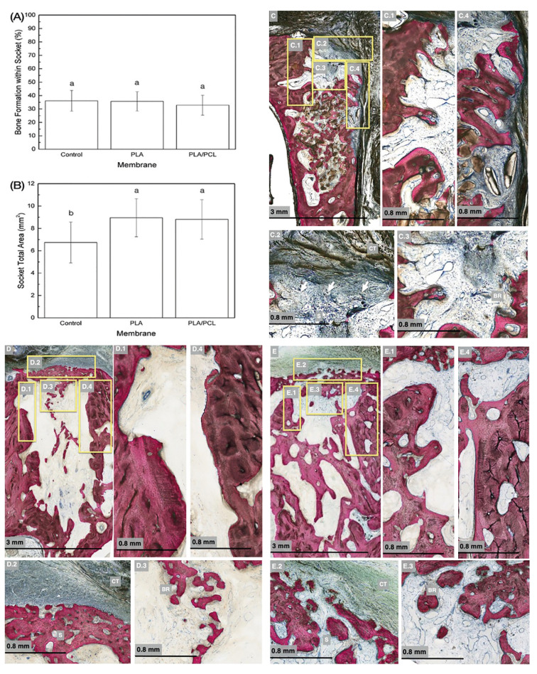
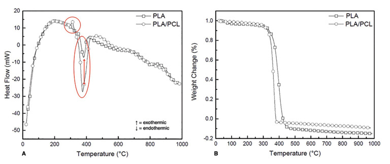
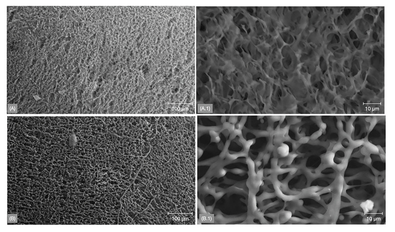
Results: Surface topography depicted a distinctive fibrous network structure for PLA membrane relative to PLA/PCL which exhibited a more porous architecture. Thermal degradation behavior/profile, observed through TGA and DSC, for both membranes was similar. Histomorphometric analysis of bone formation within the induced socket yielded 36.1 ±7.7%, 35.6 ±7.2% and 32.8 ±7.7% for control, PLA and PLA/PCL groups, respectively, with no statistically significant differences between groups (p = 0.796). Analysis of total socket area (mean ± 95% confidence intervals) yielded significantly higher values for experimental groups, PLA (8.95 ± 1.7 mm2) and PLA/PCL (8.8 ± 1.76 mm2), relative to control (6.7 ± 1.8 mm2) (p = 0.041). Residual membrane, along with mild inflammatory infiltrate was observed after the healing period irrespective of membrane type utilized.
Conclusions: Guided bone regeneration (GBR) with PLA and PLA/PCL membranes did not yield higher bone formation within the socket relative to the control group. However, an improvement in the preservation of the socket's architecture was observed.
期刊介绍:
1. Oral Medicine and Pathology:
Clinicopathological as well as medical or surgical management aspects of
diseases affecting oral mucosa, salivary glands, maxillary bones, as well as
orofacial neurological disorders, and systemic conditions with an impact on
the oral cavity.
2. Oral Surgery:
Surgical management aspects of diseases affecting oral mucosa, salivary glands,
maxillary bones, teeth, implants, oral surgical procedures. Surgical management
of diseases affecting head and neck areas.
3. Medically compromised patients in Dentistry:
Articles discussing medical problems in Odontology will also be included, with
a special focus on the clinico-odontological management of medically compromised patients, and considerations regarding high-risk or disabled patients.
4. Implantology
5. Periodontology

 求助内容:
求助内容: 应助结果提醒方式:


