Khalid Abdalla, Khaled Z Alawneh, Mohammad Al-Bdour, Abdel Qader Abu-Salih
{"title":"偏头痛和MRI:揭示潜在的关联。","authors":"Khalid Abdalla, Khaled Z Alawneh, Mohammad Al-Bdour, Abdel Qader Abu-Salih","doi":"10.1186/s13005-024-00478-2","DOIUrl":null,"url":null,"abstract":"<p><strong>Objective: </strong>This study aims to investigate the association between patients with migraine headaches and brain magnetic resonance imaging (MRI) findings.</p><p><strong>Background: </strong>Migraine is a frequently encountered primary headache disorder with a disproportionate female predominance. Diagnosis is usually based on the patient's clinical history with neuroimaging reserved for severe or atypical presentations to exclude other pathologies. Migraine patients often experience a profound impact on their quality of life.</p><p><strong>Methods: </strong>A retrospective study was conducted at King Abdullah University Hospital, Jordan, involving patients with a clinical diagnosis of migraine who had undergone MRI brain imaging between January 2021 to March 2023. Descriptive data were documented, with two independent neuro-radiologists interpreting MRI findings.</p><p><strong>Results: </strong>Our study included 670 migraine patients (510 females; mean age, 40.3 years). White matter hyperintensity lesions were found in 309 patients (46.1%), significantly affecting older age groups with a mean age of 46.8 years (p > 0.001). Additionally, gender played a role, with a higher prevalence of these lesions in female migraine patients, accounting for 79.6% (p = 0.05). Multiple logistic regression analysis proved age to be an independent risk factor for the presence of white matter hyperintensity lesions (OR: 1.0688, 95% CI: 1.0546-1.0831, p > 0.001).</p><p><strong>Conclusion: </strong>White matter hyperintensity lesions were seen in the MRI imaging of a subset of migraine patients. Patients with these lesions tend to be older and of female gender. However, the clinical significance of these findings remains unclear.</p>","PeriodicalId":12994,"journal":{"name":"Head & Face Medicine","volume":"21 1","pages":"6"},"PeriodicalIF":2.4000,"publicationDate":"2025-02-15","publicationTypes":"Journal Article","fieldsOfStudy":null,"isOpenAccess":false,"openAccessPdf":"https://www.ncbi.nlm.nih.gov/pmc/articles/PMC11830205/pdf/","citationCount":"0","resultStr":"{\"title\":\"Migraine and MRI: uncovering potential associations.\",\"authors\":\"Khalid Abdalla, Khaled Z Alawneh, Mohammad Al-Bdour, Abdel Qader Abu-Salih\",\"doi\":\"10.1186/s13005-024-00478-2\",\"DOIUrl\":null,\"url\":null,\"abstract\":\"<p><strong>Objective: </strong>This study aims to investigate the association between patients with migraine headaches and brain magnetic resonance imaging (MRI) findings.</p><p><strong>Background: </strong>Migraine is a frequently encountered primary headache disorder with a disproportionate female predominance. Diagnosis is usually based on the patient's clinical history with neuroimaging reserved for severe or atypical presentations to exclude other pathologies. Migraine patients often experience a profound impact on their quality of life.</p><p><strong>Methods: </strong>A retrospective study was conducted at King Abdullah University Hospital, Jordan, involving patients with a clinical diagnosis of migraine who had undergone MRI brain imaging between January 2021 to March 2023. Descriptive data were documented, with two independent neuro-radiologists interpreting MRI findings.</p><p><strong>Results: </strong>Our study included 670 migraine patients (510 females; mean age, 40.3 years). White matter hyperintensity lesions were found in 309 patients (46.1%), significantly affecting older age groups with a mean age of 46.8 years (p > 0.001). Additionally, gender played a role, with a higher prevalence of these lesions in female migraine patients, accounting for 79.6% (p = 0.05). Multiple logistic regression analysis proved age to be an independent risk factor for the presence of white matter hyperintensity lesions (OR: 1.0688, 95% CI: 1.0546-1.0831, p > 0.001).</p><p><strong>Conclusion: </strong>White matter hyperintensity lesions were seen in the MRI imaging of a subset of migraine patients. Patients with these lesions tend to be older and of female gender. However, the clinical significance of these findings remains unclear.</p>\",\"PeriodicalId\":12994,\"journal\":{\"name\":\"Head & Face Medicine\",\"volume\":\"21 1\",\"pages\":\"6\"},\"PeriodicalIF\":2.4000,\"publicationDate\":\"2025-02-15\",\"publicationTypes\":\"Journal Article\",\"fieldsOfStudy\":null,\"isOpenAccess\":false,\"openAccessPdf\":\"https://www.ncbi.nlm.nih.gov/pmc/articles/PMC11830205/pdf/\",\"citationCount\":\"0\",\"resultStr\":null,\"platform\":\"Semanticscholar\",\"paperid\":null,\"PeriodicalName\":\"Head & Face Medicine\",\"FirstCategoryId\":\"3\",\"ListUrlMain\":\"https://doi.org/10.1186/s13005-024-00478-2\",\"RegionNum\":2,\"RegionCategory\":\"医学\",\"ArticlePicture\":[],\"TitleCN\":null,\"AbstractTextCN\":null,\"PMCID\":null,\"EPubDate\":\"\",\"PubModel\":\"\",\"JCR\":\"Q2\",\"JCRName\":\"DENTISTRY, ORAL SURGERY & MEDICINE\",\"Score\":null,\"Total\":0}","platform":"Semanticscholar","paperid":null,"PeriodicalName":"Head & Face Medicine","FirstCategoryId":"3","ListUrlMain":"https://doi.org/10.1186/s13005-024-00478-2","RegionNum":2,"RegionCategory":"医学","ArticlePicture":[],"TitleCN":null,"AbstractTextCN":null,"PMCID":null,"EPubDate":"","PubModel":"","JCR":"Q2","JCRName":"DENTISTRY, ORAL SURGERY & MEDICINE","Score":null,"Total":0}
Migraine and MRI: uncovering potential associations.
Objective: This study aims to investigate the association between patients with migraine headaches and brain magnetic resonance imaging (MRI) findings.
Background: Migraine is a frequently encountered primary headache disorder with a disproportionate female predominance. Diagnosis is usually based on the patient's clinical history with neuroimaging reserved for severe or atypical presentations to exclude other pathologies. Migraine patients often experience a profound impact on their quality of life.
Methods: A retrospective study was conducted at King Abdullah University Hospital, Jordan, involving patients with a clinical diagnosis of migraine who had undergone MRI brain imaging between January 2021 to March 2023. Descriptive data were documented, with two independent neuro-radiologists interpreting MRI findings.
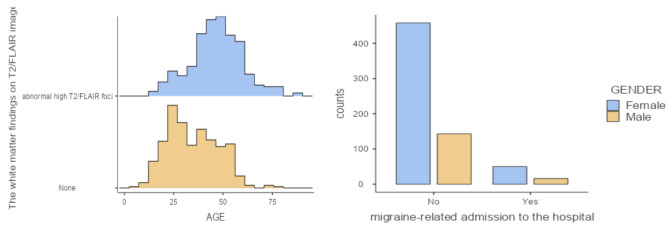
Results: Our study included 670 migraine patients (510 females; mean age, 40.3 years). White matter hyperintensity lesions were found in 309 patients (46.1%), significantly affecting older age groups with a mean age of 46.8 years (p > 0.001). Additionally, gender played a role, with a higher prevalence of these lesions in female migraine patients, accounting for 79.6% (p = 0.05). Multiple logistic regression analysis proved age to be an independent risk factor for the presence of white matter hyperintensity lesions (OR: 1.0688, 95% CI: 1.0546-1.0831, p > 0.001).
Conclusion: White matter hyperintensity lesions were seen in the MRI imaging of a subset of migraine patients. Patients with these lesions tend to be older and of female gender. However, the clinical significance of these findings remains unclear.
期刊介绍:
Head & Face Medicine is a multidisciplinary open access journal that publishes basic and clinical research concerning all aspects of cranial, facial and oral conditions.
The journal covers all aspects of cranial, facial and oral diseases and their management. It has been designed as a multidisciplinary journal for clinicians and researchers involved in the diagnostic and therapeutic aspects of diseases which affect the human head and face. The journal is wide-ranging, covering the development, aetiology, epidemiology and therapy of head and face diseases to the basic science that underlies these diseases. Management of head and face diseases includes all aspects of surgical and non-surgical treatments including psychopharmacological therapies.
 求助内容:
求助内容: 应助结果提醒方式:


