Jessica C Lal, Michelle Z Fang, Muzna Hussain, Abel Abraham, Reina Tonegawa-Kuji, Yuan Hou, Mina K Chung, Patrick Collier, Feixiong Cheng
{"title":"泛癌患者左心室心功能障碍相关血浆蛋白和代谢物的发现。","authors":"Jessica C Lal, Michelle Z Fang, Muzna Hussain, Abel Abraham, Reina Tonegawa-Kuji, Yuan Hou, Mina K Chung, Patrick Collier, Feixiong Cheng","doi":"10.1186/s40959-025-00309-6","DOIUrl":null,"url":null,"abstract":"<p><strong>Background: </strong>Cancer-therapy related cardiac dysfunction (CTRCD) remains a significant cause of morbidity and mortality in cancer survivors. In this study, we aimed to identify differential plasma proteins and metabolites associated with left ventricular dysfunction (LVD) in cancer patients.</p><p><strong>Methods: </strong>We analyzed data from 50 patients referred to the Cleveland Clinic Cardio-Oncology Center for echocardiograph assessment, integrating electronic health records, proteomic, and metabolomic profiles. LVD was defined as an ejection fraction ≤ 55% based on echocardiographic evaluation. Classification-based machine learning models were used to predict LVD using plasma metabolites and proteins as input features.</p><p><strong>Results: </strong>We identified 13 plasma proteins (P < 0.05) and 14 plasma metabolites (P < 0.05) associated with LVD. Key proteins included markers of inflammation (ST2, TNFRSF14, OPN, and AXL) and chemotaxis (RARRES2, MMP-2, MEPE, and OPN). Notably, sex-specific associations were observed, such as uridine (P = 0.003) in males. Furthermore, metabolomic features significantly associated with LVD included 1-Methyl-4-imidazoleacetic acid (P = 0.015), COL1A1 (P = 0.009), and MMP-2 (P = 0.016), and pointing to metabolic shifts and heightened inflammation in patients with LVD.</p><p><strong>Conclusion: </strong>Our findings suggest that circulating metabolites may non-invasively detect clinical and molecular differences in patients with LVD, providing insights into underlying disease pathways and potential therapeutic targets.</p>","PeriodicalId":9804,"journal":{"name":"Cardio-oncology","volume":"11 1","pages":"17"},"PeriodicalIF":3.2000,"publicationDate":"2025-02-13","publicationTypes":"Journal Article","fieldsOfStudy":null,"isOpenAccess":false,"openAccessPdf":"https://www.ncbi.nlm.nih.gov/pmc/articles/PMC11823021/pdf/","citationCount":"0","resultStr":"{\"title\":\"Discovery of plasma proteins and metabolites associated with left ventricular cardiac dysfunction in pan-cancer patients.\",\"authors\":\"Jessica C Lal, Michelle Z Fang, Muzna Hussain, Abel Abraham, Reina Tonegawa-Kuji, Yuan Hou, Mina K Chung, Patrick Collier, Feixiong Cheng\",\"doi\":\"10.1186/s40959-025-00309-6\",\"DOIUrl\":null,\"url\":null,\"abstract\":\"<p><strong>Background: </strong>Cancer-therapy related cardiac dysfunction (CTRCD) remains a significant cause of morbidity and mortality in cancer survivors. In this study, we aimed to identify differential plasma proteins and metabolites associated with left ventricular dysfunction (LVD) in cancer patients.</p><p><strong>Methods: </strong>We analyzed data from 50 patients referred to the Cleveland Clinic Cardio-Oncology Center for echocardiograph assessment, integrating electronic health records, proteomic, and metabolomic profiles. LVD was defined as an ejection fraction ≤ 55% based on echocardiographic evaluation. Classification-based machine learning models were used to predict LVD using plasma metabolites and proteins as input features.</p><p><strong>Results: </strong>We identified 13 plasma proteins (P < 0.05) and 14 plasma metabolites (P < 0.05) associated with LVD. Key proteins included markers of inflammation (ST2, TNFRSF14, OPN, and AXL) and chemotaxis (RARRES2, MMP-2, MEPE, and OPN). Notably, sex-specific associations were observed, such as uridine (P = 0.003) in males. Furthermore, metabolomic features significantly associated with LVD included 1-Methyl-4-imidazoleacetic acid (P = 0.015), COL1A1 (P = 0.009), and MMP-2 (P = 0.016), and pointing to metabolic shifts and heightened inflammation in patients with LVD.</p><p><strong>Conclusion: </strong>Our findings suggest that circulating metabolites may non-invasively detect clinical and molecular differences in patients with LVD, providing insights into underlying disease pathways and potential therapeutic targets.</p>\",\"PeriodicalId\":9804,\"journal\":{\"name\":\"Cardio-oncology\",\"volume\":\"11 1\",\"pages\":\"17\"},\"PeriodicalIF\":3.2000,\"publicationDate\":\"2025-02-13\",\"publicationTypes\":\"Journal Article\",\"fieldsOfStudy\":null,\"isOpenAccess\":false,\"openAccessPdf\":\"https://www.ncbi.nlm.nih.gov/pmc/articles/PMC11823021/pdf/\",\"citationCount\":\"0\",\"resultStr\":null,\"platform\":\"Semanticscholar\",\"paperid\":null,\"PeriodicalName\":\"Cardio-oncology\",\"FirstCategoryId\":\"1085\",\"ListUrlMain\":\"https://doi.org/10.1186/s40959-025-00309-6\",\"RegionNum\":0,\"RegionCategory\":null,\"ArticlePicture\":[],\"TitleCN\":null,\"AbstractTextCN\":null,\"PMCID\":null,\"EPubDate\":\"\",\"PubModel\":\"\",\"JCR\":\"Q2\",\"JCRName\":\"CARDIAC & CARDIOVASCULAR SYSTEMS\",\"Score\":null,\"Total\":0}","platform":"Semanticscholar","paperid":null,"PeriodicalName":"Cardio-oncology","FirstCategoryId":"1085","ListUrlMain":"https://doi.org/10.1186/s40959-025-00309-6","RegionNum":0,"RegionCategory":null,"ArticlePicture":[],"TitleCN":null,"AbstractTextCN":null,"PMCID":null,"EPubDate":"","PubModel":"","JCR":"Q2","JCRName":"CARDIAC & CARDIOVASCULAR SYSTEMS","Score":null,"Total":0}
Discovery of plasma proteins and metabolites associated with left ventricular cardiac dysfunction in pan-cancer patients.
Background: Cancer-therapy related cardiac dysfunction (CTRCD) remains a significant cause of morbidity and mortality in cancer survivors. In this study, we aimed to identify differential plasma proteins and metabolites associated with left ventricular dysfunction (LVD) in cancer patients.
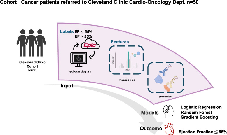
Methods: We analyzed data from 50 patients referred to the Cleveland Clinic Cardio-Oncology Center for echocardiograph assessment, integrating electronic health records, proteomic, and metabolomic profiles. LVD was defined as an ejection fraction ≤ 55% based on echocardiographic evaluation. Classification-based machine learning models were used to predict LVD using plasma metabolites and proteins as input features.
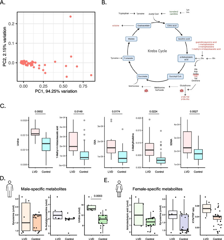
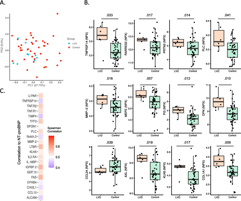
Results: We identified 13 plasma proteins (P < 0.05) and 14 plasma metabolites (P < 0.05) associated with LVD. Key proteins included markers of inflammation (ST2, TNFRSF14, OPN, and AXL) and chemotaxis (RARRES2, MMP-2, MEPE, and OPN). Notably, sex-specific associations were observed, such as uridine (P = 0.003) in males. Furthermore, metabolomic features significantly associated with LVD included 1-Methyl-4-imidazoleacetic acid (P = 0.015), COL1A1 (P = 0.009), and MMP-2 (P = 0.016), and pointing to metabolic shifts and heightened inflammation in patients with LVD.
Conclusion: Our findings suggest that circulating metabolites may non-invasively detect clinical and molecular differences in patients with LVD, providing insights into underlying disease pathways and potential therapeutic targets.

 求助内容:
求助内容: 应助结果提醒方式:


