Mahesh Bharathi, Subashini Kaliaperumal, Sandhiya Selvarajan, Renuka Srinivasan, Mary Stephen
{"title":"开角型青光眼患者24小时动态血压监测:一项横断面描述性研究。","authors":"Mahesh Bharathi, Subashini Kaliaperumal, Sandhiya Selvarajan, Renuka Srinivasan, Mary Stephen","doi":"10.22336/rjo.2024.71","DOIUrl":null,"url":null,"abstract":"<p><strong>Purpose: </strong>To compare glaucoma suspects' 24-hour blood pressure pattern with healthy subjects and the Retinal Nerve Fibre Layer (RNFL) thickness among dippers and non-dippers.</p><p><strong>Materials and methods: </strong>We included 100 patients diagnosed as glaucoma suspects in the study group and 100 age and gender-matched controls. Twenty-four-hour ambulatory blood pressure (BP) was measured using an automated BP monitoring device for mean systolic BP (SBP), mean diastolic BP (DBP), and mean arterial pressure (MAP). We classified patients into non-dippers, dippers, and overt dippers based on reduction in nocturnal MAP. Structural damage to the optic nerve head was studied by measuring the superior, inferior, and average RNFL thickness on Optical Coherence Tomography (OCT).</p><p><strong>Results: </strong>Glaucoma suspects showed lower values of day, night, and mean SBP values but higher values of day, night, and mean DBP values when compared with controls, and these were statistically significant. ANOVA and Post Hoc test (Bonferroni) analysis among glaucoma suspects showed that overt dippers had statistically significant superior, inferior, and average RNFL thinning (average 86.20 ± 12.200 µm) as compared to non-dippers and dippers (average 105 ± 11.183 and 102.19 ± 9.582 µm respectively). Pearson's correlation, used to assess the relationship between the nocturnal dip in BP and average RNFL thickness, showed a negative correlation (r = -0.396, p < 0.001).</p><p><strong>Discussion: </strong>Our study found a statistically significant decrease in systolic blood pressure day and night and an increase in diastolic blood pressure day and night in glaucoma suspects compared to normal. Mean arterial pressure did not show any significant difference, and the data obtained is comparable with previous studies. The corresponding retinal nerve fiber layer changes noted in dippers and non-dippers were also similar to those in the existing literature. This study, however, has a shortcoming of not including intraocular pressure-related nerve head changes.</p><p><strong>Conclusion: </strong>Nocturnal BP reduction was associated with structural damage to the optic nerve head in glaucoma suspects, suggesting systemic vascular etiology in the damage progression. ABP monitoring can help detect those glaucoma suspects who are mainly likely to progress so that they can be on close follow-up.</p>","PeriodicalId":94355,"journal":{"name":"Romanian journal of ophthalmology","volume":"68 4","pages":"391-397"},"PeriodicalIF":0.0000,"publicationDate":"2024-10-01","publicationTypes":"Journal Article","fieldsOfStudy":null,"isOpenAccess":false,"openAccessPdf":"https://www.ncbi.nlm.nih.gov/pmc/articles/PMC11809820/pdf/","citationCount":"0","resultStr":"{\"title\":\"Twenty-four Hour Ambulatory Blood Pressure Monitoring in Open Angle Glaucoma Suspects: A cross-sectional descriptive study.\",\"authors\":\"Mahesh Bharathi, Subashini Kaliaperumal, Sandhiya Selvarajan, Renuka Srinivasan, Mary Stephen\",\"doi\":\"10.22336/rjo.2024.71\",\"DOIUrl\":null,\"url\":null,\"abstract\":\"<p><strong>Purpose: </strong>To compare glaucoma suspects' 24-hour blood pressure pattern with healthy subjects and the Retinal Nerve Fibre Layer (RNFL) thickness among dippers and non-dippers.</p><p><strong>Materials and methods: </strong>We included 100 patients diagnosed as glaucoma suspects in the study group and 100 age and gender-matched controls. Twenty-four-hour ambulatory blood pressure (BP) was measured using an automated BP monitoring device for mean systolic BP (SBP), mean diastolic BP (DBP), and mean arterial pressure (MAP). We classified patients into non-dippers, dippers, and overt dippers based on reduction in nocturnal MAP. Structural damage to the optic nerve head was studied by measuring the superior, inferior, and average RNFL thickness on Optical Coherence Tomography (OCT).</p><p><strong>Results: </strong>Glaucoma suspects showed lower values of day, night, and mean SBP values but higher values of day, night, and mean DBP values when compared with controls, and these were statistically significant. ANOVA and Post Hoc test (Bonferroni) analysis among glaucoma suspects showed that overt dippers had statistically significant superior, inferior, and average RNFL thinning (average 86.20 ± 12.200 µm) as compared to non-dippers and dippers (average 105 ± 11.183 and 102.19 ± 9.582 µm respectively). Pearson's correlation, used to assess the relationship between the nocturnal dip in BP and average RNFL thickness, showed a negative correlation (r = -0.396, p < 0.001).</p><p><strong>Discussion: </strong>Our study found a statistically significant decrease in systolic blood pressure day and night and an increase in diastolic blood pressure day and night in glaucoma suspects compared to normal. Mean arterial pressure did not show any significant difference, and the data obtained is comparable with previous studies. The corresponding retinal nerve fiber layer changes noted in dippers and non-dippers were also similar to those in the existing literature. This study, however, has a shortcoming of not including intraocular pressure-related nerve head changes.</p><p><strong>Conclusion: </strong>Nocturnal BP reduction was associated with structural damage to the optic nerve head in glaucoma suspects, suggesting systemic vascular etiology in the damage progression. ABP monitoring can help detect those glaucoma suspects who are mainly likely to progress so that they can be on close follow-up.</p>\",\"PeriodicalId\":94355,\"journal\":{\"name\":\"Romanian journal of ophthalmology\",\"volume\":\"68 4\",\"pages\":\"391-397\"},\"PeriodicalIF\":0.0000,\"publicationDate\":\"2024-10-01\",\"publicationTypes\":\"Journal Article\",\"fieldsOfStudy\":null,\"isOpenAccess\":false,\"openAccessPdf\":\"https://www.ncbi.nlm.nih.gov/pmc/articles/PMC11809820/pdf/\",\"citationCount\":\"0\",\"resultStr\":null,\"platform\":\"Semanticscholar\",\"paperid\":null,\"PeriodicalName\":\"Romanian journal of ophthalmology\",\"FirstCategoryId\":\"1085\",\"ListUrlMain\":\"https://doi.org/10.22336/rjo.2024.71\",\"RegionNum\":0,\"RegionCategory\":null,\"ArticlePicture\":[],\"TitleCN\":null,\"AbstractTextCN\":null,\"PMCID\":null,\"EPubDate\":\"\",\"PubModel\":\"\",\"JCR\":\"\",\"JCRName\":\"\",\"Score\":null,\"Total\":0}","platform":"Semanticscholar","paperid":null,"PeriodicalName":"Romanian journal of ophthalmology","FirstCategoryId":"1085","ListUrlMain":"https://doi.org/10.22336/rjo.2024.71","RegionNum":0,"RegionCategory":null,"ArticlePicture":[],"TitleCN":null,"AbstractTextCN":null,"PMCID":null,"EPubDate":"","PubModel":"","JCR":"","JCRName":"","Score":null,"Total":0}
引用次数: 0
摘要
目的:比较青光眼疑似患者与健康人的24小时血压变化及视网膜神经纤维层(RNFL)厚度。材料和方法:我们纳入100例疑似青光眼患者作为研究组,100例年龄和性别匹配的对照组。24小时动态血压(BP)采用自动血压监测装置测量平均收缩压(SBP)、平均舒张压(DBP)和平均动脉压(MAP)。我们根据夜间MAP的减少情况将患者分为非倾斜者、倾斜者和明显倾斜者。通过光学相干断层扫描(OCT)测量上、下、平均RNFL厚度来研究视神经头的结构损伤。结果:青光眼疑似患者的日、夜、平均收缩压值低于对照组,日、夜、平均舒张压值高于对照组,差异有统计学意义。青光眼疑似患者的方差分析和事后检验(Bonferroni)分析显示,与未蘸水者和蘸水者(分别为105±11.183和102.19±9.582µm)相比,明显蘸水者的RNFL变薄(平均86.20±12.200µm)有统计学意义上的优势、不足和平均RNFL变薄(平均86.20±12.200µm)。用于评估夜间血压下降与RNFL平均厚度之间关系的Pearson相关性显示为负相关(r = -0.396, p < 0.001)。讨论:我们的研究发现,与正常患者相比,疑似青光眼患者昼夜收缩压显著降低,昼夜舒张压显著升高。平均动脉压无明显差异,所得数据与既往研究具有可比性。相应的视网膜神经纤维层变化也与已有文献相似。然而,这项研究的缺点是没有包括眼压相关的神经头变化。结论:青光眼患者夜间血压降低与视神经头结构损伤有关,提示损伤进展中有系统性血管病因。ABP监测可以帮助发现那些青光眼疑似患者,他们很可能会进展,因此可以对他们进行密切的随访。
Twenty-four Hour Ambulatory Blood Pressure Monitoring in Open Angle Glaucoma Suspects: A cross-sectional descriptive study.
Purpose: To compare glaucoma suspects' 24-hour blood pressure pattern with healthy subjects and the Retinal Nerve Fibre Layer (RNFL) thickness among dippers and non-dippers.
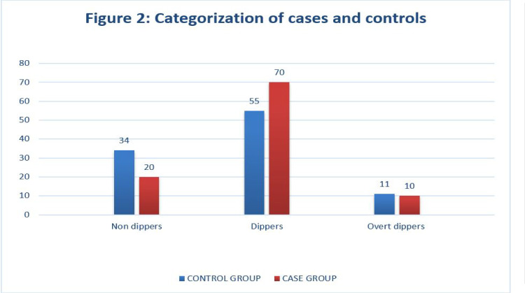
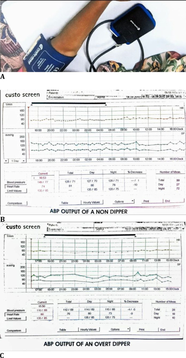
Materials and methods: We included 100 patients diagnosed as glaucoma suspects in the study group and 100 age and gender-matched controls. Twenty-four-hour ambulatory blood pressure (BP) was measured using an automated BP monitoring device for mean systolic BP (SBP), mean diastolic BP (DBP), and mean arterial pressure (MAP). We classified patients into non-dippers, dippers, and overt dippers based on reduction in nocturnal MAP. Structural damage to the optic nerve head was studied by measuring the superior, inferior, and average RNFL thickness on Optical Coherence Tomography (OCT).
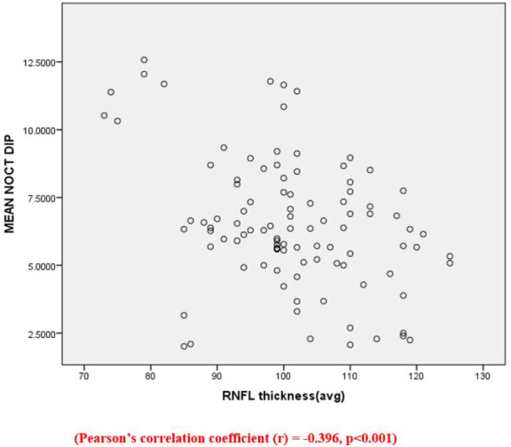
Results: Glaucoma suspects showed lower values of day, night, and mean SBP values but higher values of day, night, and mean DBP values when compared with controls, and these were statistically significant. ANOVA and Post Hoc test (Bonferroni) analysis among glaucoma suspects showed that overt dippers had statistically significant superior, inferior, and average RNFL thinning (average 86.20 ± 12.200 µm) as compared to non-dippers and dippers (average 105 ± 11.183 and 102.19 ± 9.582 µm respectively). Pearson's correlation, used to assess the relationship between the nocturnal dip in BP and average RNFL thickness, showed a negative correlation (r = -0.396, p < 0.001).
Discussion: Our study found a statistically significant decrease in systolic blood pressure day and night and an increase in diastolic blood pressure day and night in glaucoma suspects compared to normal. Mean arterial pressure did not show any significant difference, and the data obtained is comparable with previous studies. The corresponding retinal nerve fiber layer changes noted in dippers and non-dippers were also similar to those in the existing literature. This study, however, has a shortcoming of not including intraocular pressure-related nerve head changes.
Conclusion: Nocturnal BP reduction was associated with structural damage to the optic nerve head in glaucoma suspects, suggesting systemic vascular etiology in the damage progression. ABP monitoring can help detect those glaucoma suspects who are mainly likely to progress so that they can be on close follow-up.

 求助内容:
求助内容: 应助结果提醒方式:


