{"title":"乳腺癌患者胸膜内病变的鉴别诊断。","authors":"Hae Chan Song, Ho-Shin Gwak","doi":"10.14791/btrt.2025.0002","DOIUrl":null,"url":null,"abstract":"<p><p>This case introduces the differential diagnosis of a well-enhancing lesion in the prepontine cistern of a 55-year-old female patient who was diagnosed with recurrent metastatic breast cancer. The patient was diagnosed with breast cancer 11 years ago and underwent a mastectomy and subsequent adjuvant therapy. Tamoxifen had been given for 5 years, and the treatment was completed. Five years after, she found a lung nodule on her routine chest X-ray examination. Based on her past medical history, systemic cancer work-up was done and it revealed multiple lesions in T10 vertebra, lungs, and mediastinal lymph nodes. Trans-bronchial needle aspiration was performed and the biopsy was a metastatic breast cancer. Brain MRI was taken as she was complaining of headache and it showed a well-defined, ovoid enhancing 0.9-cm nodule in the right prepontine cistern. Neuro-oncology tumor board evaluated the lesion as more likely to be an asymptomatic neurogenic tumor rather than metastasis based on radiological features including brainstem surfaced location, slightly high signal intensity on T2-weighted image and no diffusion restriction. To rule out leptomeningeal metastasis, a serial cerebrospinal fluid cytology examination (×3) was done and negative for malignant cells. Follow-up brain MRIs of 2 and 9 months showed no significant changes in the pre-pontine enhancing lesion.</p>","PeriodicalId":72453,"journal":{"name":"Brain tumor research and treatment","volume":"13 1","pages":"34-38"},"PeriodicalIF":0.0000,"publicationDate":"2025-01-01","publicationTypes":"Journal Article","fieldsOfStudy":null,"isOpenAccess":false,"openAccessPdf":"https://www.ncbi.nlm.nih.gov/pmc/articles/PMC11813559/pdf/","citationCount":"0","resultStr":"{\"title\":\"Differential Diagnosis of a Well-Enhancing Intracisternal Lesion in a Breast Cancer Patient.\",\"authors\":\"Hae Chan Song, Ho-Shin Gwak\",\"doi\":\"10.14791/btrt.2025.0002\",\"DOIUrl\":null,\"url\":null,\"abstract\":\"<p><p>This case introduces the differential diagnosis of a well-enhancing lesion in the prepontine cistern of a 55-year-old female patient who was diagnosed with recurrent metastatic breast cancer. The patient was diagnosed with breast cancer 11 years ago and underwent a mastectomy and subsequent adjuvant therapy. Tamoxifen had been given for 5 years, and the treatment was completed. Five years after, she found a lung nodule on her routine chest X-ray examination. Based on her past medical history, systemic cancer work-up was done and it revealed multiple lesions in T10 vertebra, lungs, and mediastinal lymph nodes. Trans-bronchial needle aspiration was performed and the biopsy was a metastatic breast cancer. Brain MRI was taken as she was complaining of headache and it showed a well-defined, ovoid enhancing 0.9-cm nodule in the right prepontine cistern. Neuro-oncology tumor board evaluated the lesion as more likely to be an asymptomatic neurogenic tumor rather than metastasis based on radiological features including brainstem surfaced location, slightly high signal intensity on T2-weighted image and no diffusion restriction. To rule out leptomeningeal metastasis, a serial cerebrospinal fluid cytology examination (×3) was done and negative for malignant cells. Follow-up brain MRIs of 2 and 9 months showed no significant changes in the pre-pontine enhancing lesion.</p>\",\"PeriodicalId\":72453,\"journal\":{\"name\":\"Brain tumor research and treatment\",\"volume\":\"13 1\",\"pages\":\"34-38\"},\"PeriodicalIF\":0.0000,\"publicationDate\":\"2025-01-01\",\"publicationTypes\":\"Journal Article\",\"fieldsOfStudy\":null,\"isOpenAccess\":false,\"openAccessPdf\":\"https://www.ncbi.nlm.nih.gov/pmc/articles/PMC11813559/pdf/\",\"citationCount\":\"0\",\"resultStr\":null,\"platform\":\"Semanticscholar\",\"paperid\":null,\"PeriodicalName\":\"Brain tumor research and treatment\",\"FirstCategoryId\":\"1085\",\"ListUrlMain\":\"https://doi.org/10.14791/btrt.2025.0002\",\"RegionNum\":0,\"RegionCategory\":null,\"ArticlePicture\":[],\"TitleCN\":null,\"AbstractTextCN\":null,\"PMCID\":null,\"EPubDate\":\"\",\"PubModel\":\"\",\"JCR\":\"\",\"JCRName\":\"\",\"Score\":null,\"Total\":0}","platform":"Semanticscholar","paperid":null,"PeriodicalName":"Brain tumor research and treatment","FirstCategoryId":"1085","ListUrlMain":"https://doi.org/10.14791/btrt.2025.0002","RegionNum":0,"RegionCategory":null,"ArticlePicture":[],"TitleCN":null,"AbstractTextCN":null,"PMCID":null,"EPubDate":"","PubModel":"","JCR":"","JCRName":"","Score":null,"Total":0}
Differential Diagnosis of a Well-Enhancing Intracisternal Lesion in a Breast Cancer Patient.
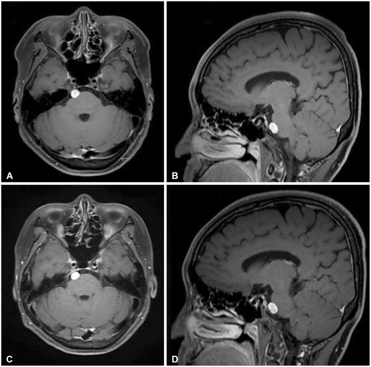
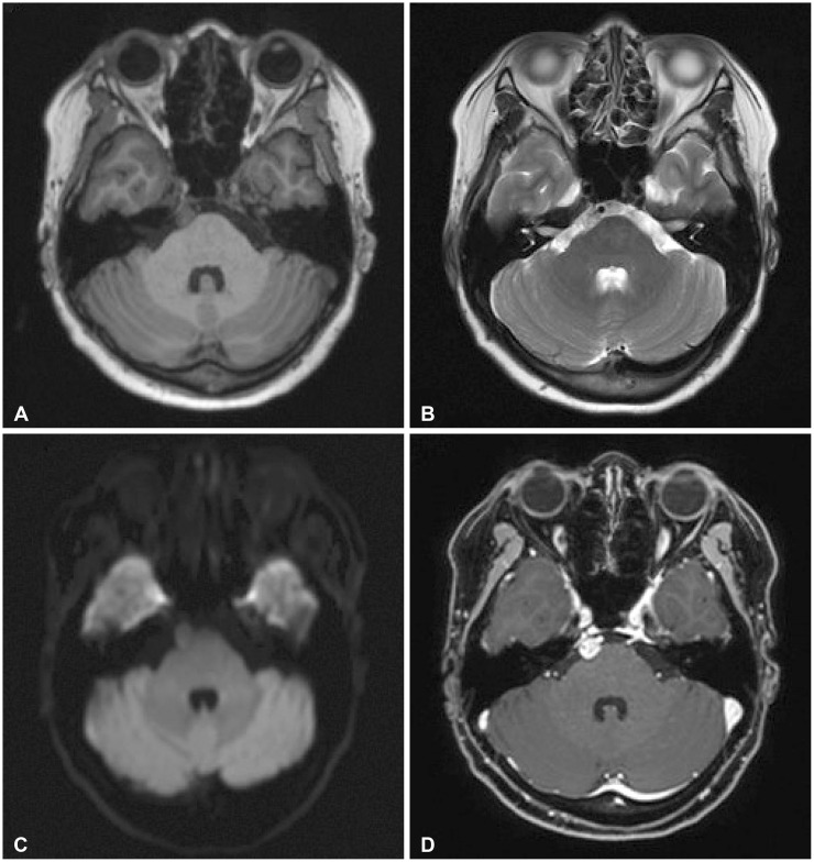
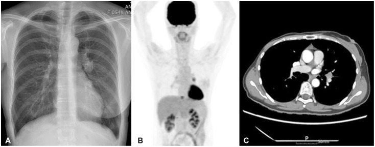
This case introduces the differential diagnosis of a well-enhancing lesion in the prepontine cistern of a 55-year-old female patient who was diagnosed with recurrent metastatic breast cancer. The patient was diagnosed with breast cancer 11 years ago and underwent a mastectomy and subsequent adjuvant therapy. Tamoxifen had been given for 5 years, and the treatment was completed. Five years after, she found a lung nodule on her routine chest X-ray examination. Based on her past medical history, systemic cancer work-up was done and it revealed multiple lesions in T10 vertebra, lungs, and mediastinal lymph nodes. Trans-bronchial needle aspiration was performed and the biopsy was a metastatic breast cancer. Brain MRI was taken as she was complaining of headache and it showed a well-defined, ovoid enhancing 0.9-cm nodule in the right prepontine cistern. Neuro-oncology tumor board evaluated the lesion as more likely to be an asymptomatic neurogenic tumor rather than metastasis based on radiological features including brainstem surfaced location, slightly high signal intensity on T2-weighted image and no diffusion restriction. To rule out leptomeningeal metastasis, a serial cerebrospinal fluid cytology examination (×3) was done and negative for malignant cells. Follow-up brain MRIs of 2 and 9 months showed no significant changes in the pre-pontine enhancing lesion.

 求助内容:
求助内容: 应助结果提醒方式:


