{"title":"可能患有阿尔茨海默病的患者的t2加权FLAIR图像上左侧梨状皮质和杏仁核的高强度与大脑皮质萎缩相关。","authors":"Hiroshi Ishizaka, Akiko Sekine, Minoru Naka, Saeki Nakano, Hiroyuki Nagase, Yoshito Tsushima","doi":"10.1177/20584601251317629","DOIUrl":null,"url":null,"abstract":"<p><strong>Background: </strong>The left piriform cortex and amygdala (PC&A) tend to be slightly hyperintense relative to the right PC&A on T2-weighted fluid-attenuated inversion recovery (T2W-FLAIR) images in patients with probable Alzheimer's disease (pAD). This likely represents the antecedent and thus advanced degeneration of the left PC&A.</p><p><strong>Purpose: </strong>To investigate the relationship between left PC&A hyperintensities and cerebral cortical atrophy on magnetic resonance (MR) voxel-based morphometry in patients with pAD and discuss how this finding could relate to AD progression.</p><p><strong>Material and methods: </strong>Patients with pAD (<i>n</i> = 47; age range = 68-93 years, mean = 80.8 ± 6.7 years; 14 men and 33 women) who underwent T2W-FLAIR imaging and MR morphometric study using a voxel-based specific regional analysis system for AD (VSRAD) were retrospectively examined. To measure signal intensity ratios of the left to right PC&A (L-PC&A/R-PC&A), regions of interest (ROIs) were set on the transaxial images in which both PC&As were most broadly depicted; the ROIs were defined as large as possible. Correlations between the L-PC&A/R-PC&A and medial temporal lobe cortical atrophy (MTLCA) as well as whole cerebral cortical atrophy (WCCA) on VSRAD were determined. Correlation between the L-PC&A/R-PC&A and age was also determined.</p><p><strong>Results: </strong>The L-PC&A/R-PC&A correlated with both MTLCA (r = 0.375, <i>p</i> = .010, 95% confidence interval [CI] = 0.095-0.600) and WCCA (r = 0.576, <i>p</i> < .001, 95% CI = 0.343-0.742). The L-PC&A/R-PC&A did not correlate with age (r = 0.013, <i>p</i> = .932, 95% CI = -0.282-0.305).</p><p><strong>Conclusion: </strong>Left-sided dominance of PC&A degeneration appeared to accelerate with the progression of AD stages.</p>","PeriodicalId":72063,"journal":{"name":"Acta radiologica open","volume":"14 2","pages":"20584601251317629"},"PeriodicalIF":1.0000,"publicationDate":"2025-02-04","publicationTypes":"Journal Article","fieldsOfStudy":null,"isOpenAccess":false,"openAccessPdf":"https://www.ncbi.nlm.nih.gov/pmc/articles/PMC11795602/pdf/","citationCount":"0","resultStr":"{\"title\":\"Hyperintensity of the left piriform cortex and amygdala on T2-weighted FLAIR images in patients with probable Alzheimer's disease correlates with cerebral cortical atrophy.\",\"authors\":\"Hiroshi Ishizaka, Akiko Sekine, Minoru Naka, Saeki Nakano, Hiroyuki Nagase, Yoshito Tsushima\",\"doi\":\"10.1177/20584601251317629\",\"DOIUrl\":null,\"url\":null,\"abstract\":\"<p><strong>Background: </strong>The left piriform cortex and amygdala (PC&A) tend to be slightly hyperintense relative to the right PC&A on T2-weighted fluid-attenuated inversion recovery (T2W-FLAIR) images in patients with probable Alzheimer's disease (pAD). This likely represents the antecedent and thus advanced degeneration of the left PC&A.</p><p><strong>Purpose: </strong>To investigate the relationship between left PC&A hyperintensities and cerebral cortical atrophy on magnetic resonance (MR) voxel-based morphometry in patients with pAD and discuss how this finding could relate to AD progression.</p><p><strong>Material and methods: </strong>Patients with pAD (<i>n</i> = 47; age range = 68-93 years, mean = 80.8 ± 6.7 years; 14 men and 33 women) who underwent T2W-FLAIR imaging and MR morphometric study using a voxel-based specific regional analysis system for AD (VSRAD) were retrospectively examined. To measure signal intensity ratios of the left to right PC&A (L-PC&A/R-PC&A), regions of interest (ROIs) were set on the transaxial images in which both PC&As were most broadly depicted; the ROIs were defined as large as possible. Correlations between the L-PC&A/R-PC&A and medial temporal lobe cortical atrophy (MTLCA) as well as whole cerebral cortical atrophy (WCCA) on VSRAD were determined. Correlation between the L-PC&A/R-PC&A and age was also determined.</p><p><strong>Results: </strong>The L-PC&A/R-PC&A correlated with both MTLCA (r = 0.375, <i>p</i> = .010, 95% confidence interval [CI] = 0.095-0.600) and WCCA (r = 0.576, <i>p</i> < .001, 95% CI = 0.343-0.742). The L-PC&A/R-PC&A did not correlate with age (r = 0.013, <i>p</i> = .932, 95% CI = -0.282-0.305).</p><p><strong>Conclusion: </strong>Left-sided dominance of PC&A degeneration appeared to accelerate with the progression of AD stages.</p>\",\"PeriodicalId\":72063,\"journal\":{\"name\":\"Acta radiologica open\",\"volume\":\"14 2\",\"pages\":\"20584601251317629\"},\"PeriodicalIF\":1.0000,\"publicationDate\":\"2025-02-04\",\"publicationTypes\":\"Journal Article\",\"fieldsOfStudy\":null,\"isOpenAccess\":false,\"openAccessPdf\":\"https://www.ncbi.nlm.nih.gov/pmc/articles/PMC11795602/pdf/\",\"citationCount\":\"0\",\"resultStr\":null,\"platform\":\"Semanticscholar\",\"paperid\":null,\"PeriodicalName\":\"Acta radiologica open\",\"FirstCategoryId\":\"1085\",\"ListUrlMain\":\"https://doi.org/10.1177/20584601251317629\",\"RegionNum\":0,\"RegionCategory\":null,\"ArticlePicture\":[],\"TitleCN\":null,\"AbstractTextCN\":null,\"PMCID\":null,\"EPubDate\":\"2025/2/1 0:00:00\",\"PubModel\":\"eCollection\",\"JCR\":\"Q4\",\"JCRName\":\"RADIOLOGY, NUCLEAR MEDICINE & MEDICAL IMAGING\",\"Score\":null,\"Total\":0}","platform":"Semanticscholar","paperid":null,"PeriodicalName":"Acta radiologica open","FirstCategoryId":"1085","ListUrlMain":"https://doi.org/10.1177/20584601251317629","RegionNum":0,"RegionCategory":null,"ArticlePicture":[],"TitleCN":null,"AbstractTextCN":null,"PMCID":null,"EPubDate":"2025/2/1 0:00:00","PubModel":"eCollection","JCR":"Q4","JCRName":"RADIOLOGY, NUCLEAR MEDICINE & MEDICAL IMAGING","Score":null,"Total":0}
引用次数: 0
摘要
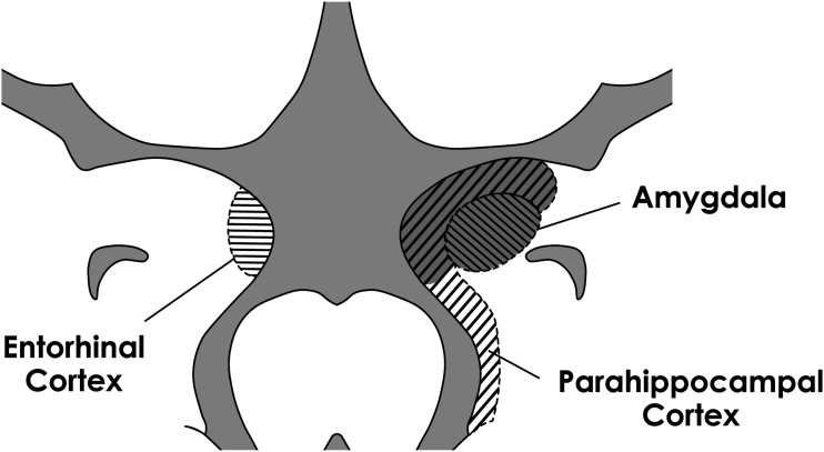
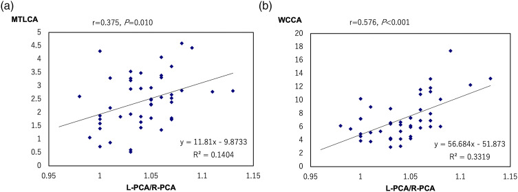
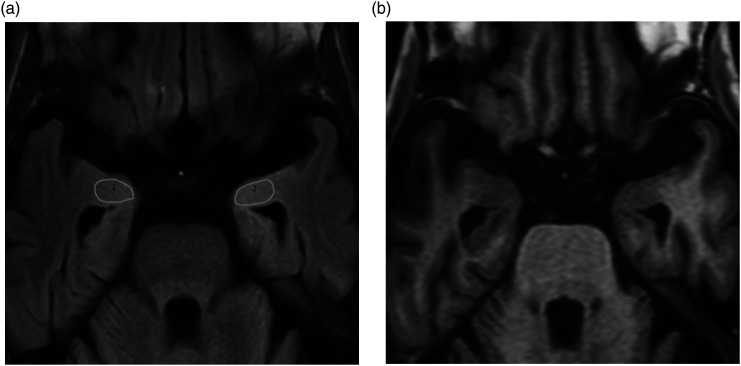
背景:在疑似阿尔茨海默病(pAD)患者的t2加权液体衰减反转恢复(T2W-FLAIR)图像上,左侧梨状皮质和杏仁核(PC&A)相对于右侧PC&A呈轻微高信号。这可能代表了左侧PC&A的先前和晚期退变。目的:通过磁共振体素形态学分析研究pAD患者左PC&A高信号与大脑皮层萎缩的关系,并探讨这一发现与AD进展的关系。材料和方法:pAD患者(n = 47;年龄范围= 68 ~ 93岁,平均= 80.8±6.7岁;采用基于体素的AD特异性区域分析系统(VSRAD)对14名男性和33名女性进行了T2W-FLAIR成像和MR形态计量学研究。为了测量左右PC&A的信号强度比(L-PC&A/R-PC&A),在两个PC&A最广泛描绘的跨轴图像上设置感兴趣区域(roi);投资回报率被定义得尽可能大。测定L-PC&A/R-PC&A与VSRAD患者内侧颞叶皮质萎缩(MTLCA)及全脑皮质萎缩(WCCA)的相关性。L-PC&A/R-PC&A与年龄的相关性也被确定。结果:L-PC&A/ r - pc&a与MTLCA (r = 0.375, p = 0.010, 95%可信区间[CI] = 0.095 ~ 0.600)和WCCA (r = 0.576, p < 0.001, 95% CI = 0.343 ~ 0.742)均相关。L-PC&A/ r - pc&a与年龄无相关性(r = 0.013, p = 0.932, 95% CI = -0.282 ~ 0.305)。结论:随着AD分期的进展,PC&A左侧优势变性表现为加速。
Hyperintensity of the left piriform cortex and amygdala on T2-weighted FLAIR images in patients with probable Alzheimer's disease correlates with cerebral cortical atrophy.
Background: The left piriform cortex and amygdala (PC&A) tend to be slightly hyperintense relative to the right PC&A on T2-weighted fluid-attenuated inversion recovery (T2W-FLAIR) images in patients with probable Alzheimer's disease (pAD). This likely represents the antecedent and thus advanced degeneration of the left PC&A.
Purpose: To investigate the relationship between left PC&A hyperintensities and cerebral cortical atrophy on magnetic resonance (MR) voxel-based morphometry in patients with pAD and discuss how this finding could relate to AD progression.
Material and methods: Patients with pAD (n = 47; age range = 68-93 years, mean = 80.8 ± 6.7 years; 14 men and 33 women) who underwent T2W-FLAIR imaging and MR morphometric study using a voxel-based specific regional analysis system for AD (VSRAD) were retrospectively examined. To measure signal intensity ratios of the left to right PC&A (L-PC&A/R-PC&A), regions of interest (ROIs) were set on the transaxial images in which both PC&As were most broadly depicted; the ROIs were defined as large as possible. Correlations between the L-PC&A/R-PC&A and medial temporal lobe cortical atrophy (MTLCA) as well as whole cerebral cortical atrophy (WCCA) on VSRAD were determined. Correlation between the L-PC&A/R-PC&A and age was also determined.
Results: The L-PC&A/R-PC&A correlated with both MTLCA (r = 0.375, p = .010, 95% confidence interval [CI] = 0.095-0.600) and WCCA (r = 0.576, p < .001, 95% CI = 0.343-0.742). The L-PC&A/R-PC&A did not correlate with age (r = 0.013, p = .932, 95% CI = -0.282-0.305).
Conclusion: Left-sided dominance of PC&A degeneration appeared to accelerate with the progression of AD stages.

 求助内容:
求助内容: 应助结果提醒方式:


