Mahdi Mohammadi, Shahram Samadi, Seyed Amir Hossein Batouli, Khalil Pestei, Mohammad Ali Oghabian
{"title":"降低氧提取分数作为阻塞性睡眠呼吸暂停患者认知缺陷的生物标志物","authors":"Mahdi Mohammadi, Shahram Samadi, Seyed Amir Hossein Batouli, Khalil Pestei, Mohammad Ali Oghabian","doi":"10.1002/brb3.70273","DOIUrl":null,"url":null,"abstract":"<div>\n \n \n <section>\n \n <h3> Background</h3>\n \n <p>Obstructive sleep apnea (OSA) is characterized by disruptive breathing, resulting in a decline in cognitive performance. This study investigates the role of oxygen extraction fraction (OEF) and quantitative susceptibility mapping (QSM) in OSA-related cognitive impairment.</p>\n </section>\n \n <section>\n \n <h3> Methods</h3>\n \n <p>The study recruited 15 patients with confirmed OSA and 16 healthy controls, who underwent overnight polysomnography and brain MRI using a 3 Tesla machine and 64-channel head coil. A two-step MRI analysis was employed to measure OEF. QSM was first created by processing separate phase and magnitude images. OEF maps were then generated by identifying veins based on their susceptibility. Volumetric analysis was performed using the FreeSurfer. Neuropsychological tests were administered to evaluate cognition.</p>\n </section>\n \n <section>\n \n <h3> Results</h3>\n \n <p>The analysis of OEF revealed significantly lower values in various cerebral cortical regions of OSA patients than in controls. Notably, OEF in the cerebral cortex and frontal, temporal, and occipital regions showed negative correlations with the duration of stage N2 sleep (highest correlation between N2 and right temporal OEF: <i>p</i> = 0.005, <i>r</i> = −0.681). Furthermore, poorer performance on neuropsychological tests, such as the backward digit span test, was significantly correlated with reduced OEF in the left hemisphere (<i>p</i> = 0.016), left cerebral cortex (<i>p</i> = 0.019), right frontal (<i>p</i> = 0.034), left frontal (<i>p</i> = 0.014), left parietal (<i>p</i> = 0.008), left temporal (<i>p</i> = 0.048), and left occipital lobes (<i>p</i> = 0.015). No significant differences in QSM or brain volume were observed.</p>\n </section>\n \n <section>\n \n <h3> Conclusions</h3>\n \n <p>Decreased OEF emerges as a potential biomarker for cognitive deficits in OSA, suggesting disturbances in cerebral oxygen metabolism may underlie cognitive impairments. These findings underscore the importance of investigating physiological markers in understanding OSA-related cognitive dysfunction.</p>\n </section>\n </div>","PeriodicalId":9081,"journal":{"name":"Brain and Behavior","volume":"15 2","pages":""},"PeriodicalIF":2.7000,"publicationDate":"2025-02-06","publicationTypes":"Journal Article","fieldsOfStudy":null,"isOpenAccess":false,"openAccessPdf":"https://onlinelibrary.wiley.com/doi/epdf/10.1002/brb3.70273","citationCount":"0","resultStr":"{\"title\":\"Reduced Oxygen Extraction Fraction as a Biomarker for Cognitive Deficits in Obstructive Sleep Apnea\",\"authors\":\"Mahdi Mohammadi, Shahram Samadi, Seyed Amir Hossein Batouli, Khalil Pestei, Mohammad Ali Oghabian\",\"doi\":\"10.1002/brb3.70273\",\"DOIUrl\":null,\"url\":null,\"abstract\":\"<div>\\n \\n \\n <section>\\n \\n <h3> Background</h3>\\n \\n <p>Obstructive sleep apnea (OSA) is characterized by disruptive breathing, resulting in a decline in cognitive performance. This study investigates the role of oxygen extraction fraction (OEF) and quantitative susceptibility mapping (QSM) in OSA-related cognitive impairment.</p>\\n </section>\\n \\n <section>\\n \\n <h3> Methods</h3>\\n \\n <p>The study recruited 15 patients with confirmed OSA and 16 healthy controls, who underwent overnight polysomnography and brain MRI using a 3 Tesla machine and 64-channel head coil. A two-step MRI analysis was employed to measure OEF. QSM was first created by processing separate phase and magnitude images. OEF maps were then generated by identifying veins based on their susceptibility. Volumetric analysis was performed using the FreeSurfer. Neuropsychological tests were administered to evaluate cognition.</p>\\n </section>\\n \\n <section>\\n \\n <h3> Results</h3>\\n \\n <p>The analysis of OEF revealed significantly lower values in various cerebral cortical regions of OSA patients than in controls. Notably, OEF in the cerebral cortex and frontal, temporal, and occipital regions showed negative correlations with the duration of stage N2 sleep (highest correlation between N2 and right temporal OEF: <i>p</i> = 0.005, <i>r</i> = −0.681). Furthermore, poorer performance on neuropsychological tests, such as the backward digit span test, was significantly correlated with reduced OEF in the left hemisphere (<i>p</i> = 0.016), left cerebral cortex (<i>p</i> = 0.019), right frontal (<i>p</i> = 0.034), left frontal (<i>p</i> = 0.014), left parietal (<i>p</i> = 0.008), left temporal (<i>p</i> = 0.048), and left occipital lobes (<i>p</i> = 0.015). No significant differences in QSM or brain volume were observed.</p>\\n </section>\\n \\n <section>\\n \\n <h3> Conclusions</h3>\\n \\n <p>Decreased OEF emerges as a potential biomarker for cognitive deficits in OSA, suggesting disturbances in cerebral oxygen metabolism may underlie cognitive impairments. These findings underscore the importance of investigating physiological markers in understanding OSA-related cognitive dysfunction.</p>\\n </section>\\n </div>\",\"PeriodicalId\":9081,\"journal\":{\"name\":\"Brain and Behavior\",\"volume\":\"15 2\",\"pages\":\"\"},\"PeriodicalIF\":2.7000,\"publicationDate\":\"2025-02-06\",\"publicationTypes\":\"Journal Article\",\"fieldsOfStudy\":null,\"isOpenAccess\":false,\"openAccessPdf\":\"https://onlinelibrary.wiley.com/doi/epdf/10.1002/brb3.70273\",\"citationCount\":\"0\",\"resultStr\":null,\"platform\":\"Semanticscholar\",\"paperid\":null,\"PeriodicalName\":\"Brain and Behavior\",\"FirstCategoryId\":\"102\",\"ListUrlMain\":\"https://onlinelibrary.wiley.com/doi/10.1002/brb3.70273\",\"RegionNum\":3,\"RegionCategory\":\"心理学\",\"ArticlePicture\":[],\"TitleCN\":null,\"AbstractTextCN\":null,\"PMCID\":null,\"EPubDate\":\"\",\"PubModel\":\"\",\"JCR\":\"Q2\",\"JCRName\":\"BEHAVIORAL SCIENCES\",\"Score\":null,\"Total\":0}","platform":"Semanticscholar","paperid":null,"PeriodicalName":"Brain and Behavior","FirstCategoryId":"102","ListUrlMain":"https://onlinelibrary.wiley.com/doi/10.1002/brb3.70273","RegionNum":3,"RegionCategory":"心理学","ArticlePicture":[],"TitleCN":null,"AbstractTextCN":null,"PMCID":null,"EPubDate":"","PubModel":"","JCR":"Q2","JCRName":"BEHAVIORAL SCIENCES","Score":null,"Total":0}
Reduced Oxygen Extraction Fraction as a Biomarker for Cognitive Deficits in Obstructive Sleep Apnea
Background
Obstructive sleep apnea (OSA) is characterized by disruptive breathing, resulting in a decline in cognitive performance. This study investigates the role of oxygen extraction fraction (OEF) and quantitative susceptibility mapping (QSM) in OSA-related cognitive impairment.
Methods
The study recruited 15 patients with confirmed OSA and 16 healthy controls, who underwent overnight polysomnography and brain MRI using a 3 Tesla machine and 64-channel head coil. A two-step MRI analysis was employed to measure OEF. QSM was first created by processing separate phase and magnitude images. OEF maps were then generated by identifying veins based on their susceptibility. Volumetric analysis was performed using the FreeSurfer. Neuropsychological tests were administered to evaluate cognition.
Results
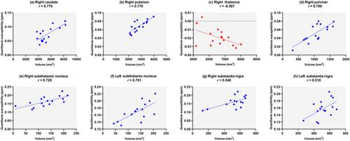
The analysis of OEF revealed significantly lower values in various cerebral cortical regions of OSA patients than in controls. Notably, OEF in the cerebral cortex and frontal, temporal, and occipital regions showed negative correlations with the duration of stage N2 sleep (highest correlation between N2 and right temporal OEF: p = 0.005, r = −0.681). Furthermore, poorer performance on neuropsychological tests, such as the backward digit span test, was significantly correlated with reduced OEF in the left hemisphere (p = 0.016), left cerebral cortex (p = 0.019), right frontal (p = 0.034), left frontal (p = 0.014), left parietal (p = 0.008), left temporal (p = 0.048), and left occipital lobes (p = 0.015). No significant differences in QSM or brain volume were observed.
Conclusions
Decreased OEF emerges as a potential biomarker for cognitive deficits in OSA, suggesting disturbances in cerebral oxygen metabolism may underlie cognitive impairments. These findings underscore the importance of investigating physiological markers in understanding OSA-related cognitive dysfunction.
期刊介绍:
Brain and Behavior is supported by other journals published by Wiley, including a number of society-owned journals. The journals listed below support Brain and Behavior and participate in the Manuscript Transfer Program by referring articles of suitable quality and offering authors the option to have their paper, with any peer review reports, automatically transferred to Brain and Behavior.
* [Acta Psychiatrica Scandinavica](https://publons.com/journal/1366/acta-psychiatrica-scandinavica)
* [Addiction Biology](https://publons.com/journal/1523/addiction-biology)
* [Aggressive Behavior](https://publons.com/journal/3611/aggressive-behavior)
* [Brain Pathology](https://publons.com/journal/1787/brain-pathology)
* [Child: Care, Health and Development](https://publons.com/journal/6111/child-care-health-and-development)
* [Criminal Behaviour and Mental Health](https://publons.com/journal/3839/criminal-behaviour-and-mental-health)
* [Depression and Anxiety](https://publons.com/journal/1528/depression-and-anxiety)
* Developmental Neurobiology
* [Developmental Science](https://publons.com/journal/1069/developmental-science)
* [European Journal of Neuroscience](https://publons.com/journal/1441/european-journal-of-neuroscience)
* [Genes, Brain and Behavior](https://publons.com/journal/1635/genes-brain-and-behavior)
* [GLIA](https://publons.com/journal/1287/glia)
* [Hippocampus](https://publons.com/journal/1056/hippocampus)
* [Human Brain Mapping](https://publons.com/journal/500/human-brain-mapping)
* [Journal for the Theory of Social Behaviour](https://publons.com/journal/7330/journal-for-the-theory-of-social-behaviour)
* [Journal of Comparative Neurology](https://publons.com/journal/1306/journal-of-comparative-neurology)
* [Journal of Neuroimaging](https://publons.com/journal/6379/journal-of-neuroimaging)
* [Journal of Neuroscience Research](https://publons.com/journal/2778/journal-of-neuroscience-research)
* [Journal of Organizational Behavior](https://publons.com/journal/1123/journal-of-organizational-behavior)
* [Journal of the Peripheral Nervous System](https://publons.com/journal/3929/journal-of-the-peripheral-nervous-system)
* [Muscle & Nerve](https://publons.com/journal/4448/muscle-and-nerve)
* [Neural Pathology and Applied Neurobiology](https://publons.com/journal/2401/neuropathology-and-applied-neurobiology)
 求助内容:
求助内容: 应助结果提醒方式:


