Davide Greco, Alexander Drakew, Nina Rößler, Tassilo Jungenitz, Peter Jedlicka, Thomas Deller
{"title":"内嗅损伤后小鼠齿状回颗粒细胞的延时成像揭示了细胞对去神经控制的异质反应。","authors":"Davide Greco, Alexander Drakew, Nina Rößler, Tassilo Jungenitz, Peter Jedlicka, Thomas Deller","doi":"10.3389/fnana.2024.1513511","DOIUrl":null,"url":null,"abstract":"<p><p>Denervation of neurons is a network consequence of brain injury. The effects of denervation on neurons can be readily studied <i>in vitro</i> using organotypic slice cultures of entorhinal cortex and hippocampus. Following transection of the entorhino-dentate projection, granule cells (GCs) are denervated and show on average a transient loss of spines on their denervated distal dendrites but not on their non-denervated proximal dendrites. In the present study, we addressed the question how single GCs and their denervated and non-denervated segments react to entorhinal denervation. Local adeno-associated virus (AAV)-injections were employed to transduce dentate GCs with tdTomato and entorhinal projection neurons with EGFP. This made it possible to visualize both innervating entorhinal fibers and their target neurons and to identify dendritic segments located in the \"entorhinal\" and the \"hippocampal\" zone of the dentate gyrus. Confocal time-lapse imaging was used to image distal and proximal segments of single GCs after entorhinal denervation. Time-matched non-denervated cultures served as controls. In line with previous reports, average dendritic spine loss was ~30% (2-4 days post-lesion) in the denervated zone. However, individual GCs showed considerable variability in their response to denervation in both layers, and both decreases as well as increases in spine density were observed at the single cell level. Based on the standard deviations and the effect sizes observed in this study, a computer simulation yielded recommendations for the minimum number of neurons that should be analyzed in future studies using the entorhinal <i>in vitro</i> denervation model.</p>","PeriodicalId":12572,"journal":{"name":"Frontiers in Neuroanatomy","volume":"18 ","pages":"1513511"},"PeriodicalIF":2.3000,"publicationDate":"2025-01-21","publicationTypes":"Journal Article","fieldsOfStudy":null,"isOpenAccess":false,"openAccessPdf":"https://www.ncbi.nlm.nih.gov/pmc/articles/PMC11790675/pdf/","citationCount":"0","resultStr":"{\"title\":\"Time-lapse imaging of identified granule cells in the mouse dentate gyrus after entorhinal lesion <i>in vitro</i> reveals heterogeneous cellular responses to denervation.\",\"authors\":\"Davide Greco, Alexander Drakew, Nina Rößler, Tassilo Jungenitz, Peter Jedlicka, Thomas Deller\",\"doi\":\"10.3389/fnana.2024.1513511\",\"DOIUrl\":null,\"url\":null,\"abstract\":\"<p><p>Denervation of neurons is a network consequence of brain injury. The effects of denervation on neurons can be readily studied <i>in vitro</i> using organotypic slice cultures of entorhinal cortex and hippocampus. Following transection of the entorhino-dentate projection, granule cells (GCs) are denervated and show on average a transient loss of spines on their denervated distal dendrites but not on their non-denervated proximal dendrites. In the present study, we addressed the question how single GCs and their denervated and non-denervated segments react to entorhinal denervation. Local adeno-associated virus (AAV)-injections were employed to transduce dentate GCs with tdTomato and entorhinal projection neurons with EGFP. This made it possible to visualize both innervating entorhinal fibers and their target neurons and to identify dendritic segments located in the \\\"entorhinal\\\" and the \\\"hippocampal\\\" zone of the dentate gyrus. Confocal time-lapse imaging was used to image distal and proximal segments of single GCs after entorhinal denervation. Time-matched non-denervated cultures served as controls. In line with previous reports, average dendritic spine loss was ~30% (2-4 days post-lesion) in the denervated zone. However, individual GCs showed considerable variability in their response to denervation in both layers, and both decreases as well as increases in spine density were observed at the single cell level. Based on the standard deviations and the effect sizes observed in this study, a computer simulation yielded recommendations for the minimum number of neurons that should be analyzed in future studies using the entorhinal <i>in vitro</i> denervation model.</p>\",\"PeriodicalId\":12572,\"journal\":{\"name\":\"Frontiers in Neuroanatomy\",\"volume\":\"18 \",\"pages\":\"1513511\"},\"PeriodicalIF\":2.3000,\"publicationDate\":\"2025-01-21\",\"publicationTypes\":\"Journal Article\",\"fieldsOfStudy\":null,\"isOpenAccess\":false,\"openAccessPdf\":\"https://www.ncbi.nlm.nih.gov/pmc/articles/PMC11790675/pdf/\",\"citationCount\":\"0\",\"resultStr\":null,\"platform\":\"Semanticscholar\",\"paperid\":null,\"PeriodicalName\":\"Frontiers in Neuroanatomy\",\"FirstCategoryId\":\"3\",\"ListUrlMain\":\"https://doi.org/10.3389/fnana.2024.1513511\",\"RegionNum\":4,\"RegionCategory\":\"医学\",\"ArticlePicture\":[],\"TitleCN\":null,\"AbstractTextCN\":null,\"PMCID\":null,\"EPubDate\":\"2024/1/1 0:00:00\",\"PubModel\":\"eCollection\",\"JCR\":\"Q1\",\"JCRName\":\"ANATOMY & MORPHOLOGY\",\"Score\":null,\"Total\":0}","platform":"Semanticscholar","paperid":null,"PeriodicalName":"Frontiers in Neuroanatomy","FirstCategoryId":"3","ListUrlMain":"https://doi.org/10.3389/fnana.2024.1513511","RegionNum":4,"RegionCategory":"医学","ArticlePicture":[],"TitleCN":null,"AbstractTextCN":null,"PMCID":null,"EPubDate":"2024/1/1 0:00:00","PubModel":"eCollection","JCR":"Q1","JCRName":"ANATOMY & MORPHOLOGY","Score":null,"Total":0}
Time-lapse imaging of identified granule cells in the mouse dentate gyrus after entorhinal lesion in vitro reveals heterogeneous cellular responses to denervation.
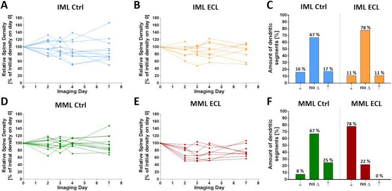
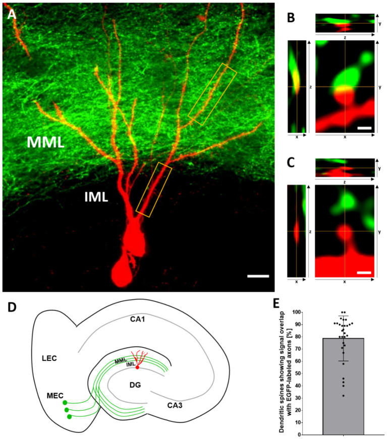
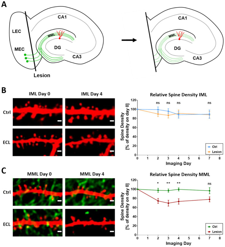
Denervation of neurons is a network consequence of brain injury. The effects of denervation on neurons can be readily studied in vitro using organotypic slice cultures of entorhinal cortex and hippocampus. Following transection of the entorhino-dentate projection, granule cells (GCs) are denervated and show on average a transient loss of spines on their denervated distal dendrites but not on their non-denervated proximal dendrites. In the present study, we addressed the question how single GCs and their denervated and non-denervated segments react to entorhinal denervation. Local adeno-associated virus (AAV)-injections were employed to transduce dentate GCs with tdTomato and entorhinal projection neurons with EGFP. This made it possible to visualize both innervating entorhinal fibers and their target neurons and to identify dendritic segments located in the "entorhinal" and the "hippocampal" zone of the dentate gyrus. Confocal time-lapse imaging was used to image distal and proximal segments of single GCs after entorhinal denervation. Time-matched non-denervated cultures served as controls. In line with previous reports, average dendritic spine loss was ~30% (2-4 days post-lesion) in the denervated zone. However, individual GCs showed considerable variability in their response to denervation in both layers, and both decreases as well as increases in spine density were observed at the single cell level. Based on the standard deviations and the effect sizes observed in this study, a computer simulation yielded recommendations for the minimum number of neurons that should be analyzed in future studies using the entorhinal in vitro denervation model.
期刊介绍:
Frontiers in Neuroanatomy publishes rigorously peer-reviewed research revealing important aspects of the anatomical organization of all nervous systems across all species. Specialty Chief Editor Javier DeFelipe at the Cajal Institute (CSIC) is supported by an outstanding Editorial Board of international experts. This multidisciplinary open-access journal is at the forefront of disseminating and communicating scientific knowledge and impactful discoveries to researchers, academics, clinicians and the public worldwide.

 求助内容:
求助内容: 应助结果提醒方式:


